Super-resolution for localizing electrode grids as small, deformable objects during epilepsy surgery using augmented reality headsets - Report - MDSpire
Advertisement
Super-resolution for localizing electrode grids as small, deformable objects during epilepsy surgery using augmented reality headsets
Enhanced Localization of Flexible Electrode Grids in Epilepsy Surgery Using AR and AI
Overview
This study introduces a novel method combining augmented reality head-mounted displays (AR HMDs) with artificial intelligence (AI) to improve the localization of intraoperative electrocorticography (ioECoG) grids in epilepsy surgery. The approach enhances accuracy and workflow efficiency compared to traditional photograph-based methods, enabling real-time visualization and precise electrode detection.
Background
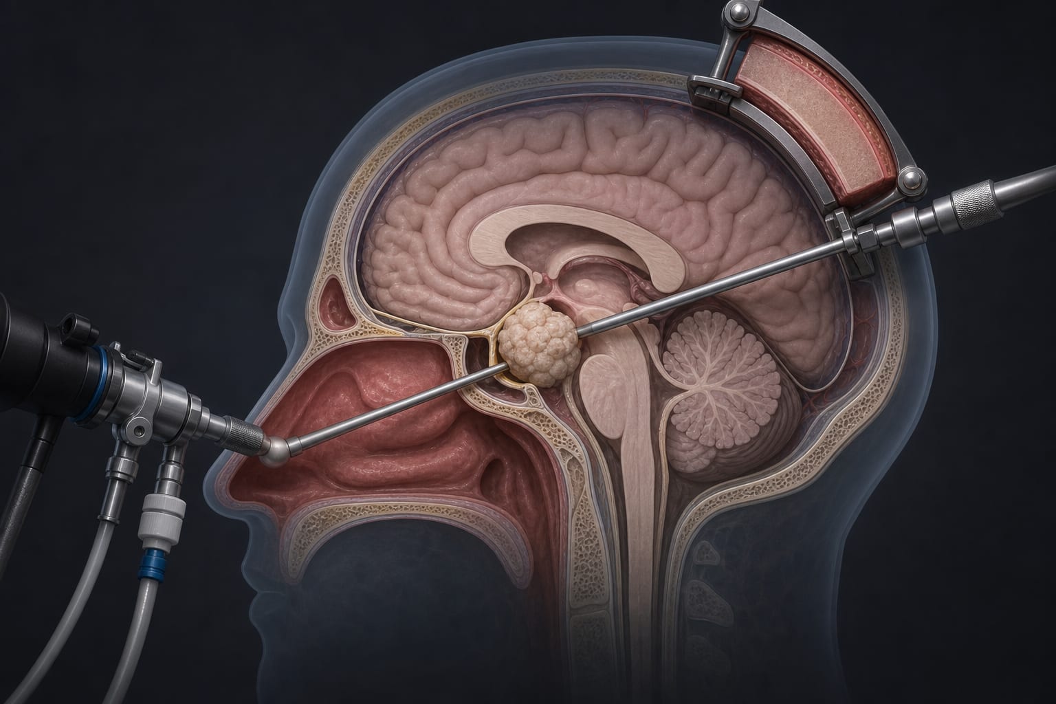
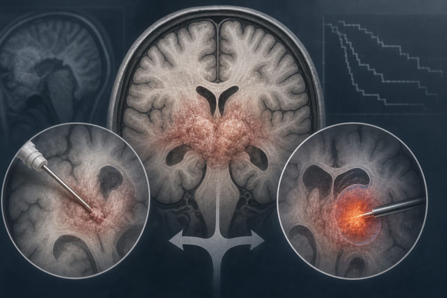
Epilepsy affects approximately 50 million people worldwide, with about 30% experiencing seizures refractory to medication. Surgical treatment for focal epilepsy relies on precise localization and resection of epileptic tissue, often guided by ioECoG grids placed on the cortex. Current localization techniques depend on visual inspection or photograph-based alignment, which are time-consuming, require expertise, and are limited by grid visibility and deformation. Augmented reality (AR) and AI technologies offer promising solutions to overcome these challenges by providing real-time, accurate, and hands-free localization.
Data Highlights
The proposed system uses a 64-electrode ioECoG grid with 5 mm inter-electrode spacing. The method involves wireless transmission of stereo images from AR HMD cameras, image undistortion and rectification, bounding box estimation, super-resolution upscaling by a factor of four, and 2D pose estimation to detect electrode centers. Prior methods achieved mean localization errors around 2.0 mm with maximum errors up to 6.4 mm, but required hours and specialized expertise. The new approach aims to maintain accuracy within clinically acceptable thresholds (<5 mm) while improving speed and usability.
Key Findings
Traditional ioECoG grid localization methods rely on visual inspection or photograph-based alignment, which are time-consuming and limited by grid occlusion.
AR HMDs provide hands-free, real-time visualization and localization capabilities, integrating seamlessly into surgical workflows.
AI techniques, including deep learning for object detection, super-resolution, and 2D pose estimation, can overcome challenges posed by low-resolution sensors and deformable grids.
The proposed AR and AI-based method enables automatic detection of individual electrode centers with improved accuracy and speed compared to manual methods.
Use of synthetic data addresses the scarcity of annotated medical images for training AI models in this application.
Comparison with a commercial NDI tracking system as ground truth demonstrates the feasibility and potential clinical utility of the approach.
Clinical Implications
Integrating AR HMDs with AI-based localization can enhance surgical precision by providing accurate, real-time electrode grid positioning without disrupting workflow. This technology may reduce operative time and reliance on specialized expertise, potentially improving outcomes in epilepsy surgery. Adoption of such systems could facilitate more complete resection of epileptic tissue, thereby increasing the likelihood of seizure freedom.
Conclusion
The combination of augmented reality and artificial intelligence represents a promising advancement for intraoperative localization of flexible electrode grids in epilepsy surgery. This approach improves accuracy and efficiency, supporting better surgical decision-making and patient outcomes.
References
Pieters et al. -- Comparison of ioECoG Grid Localization Methods