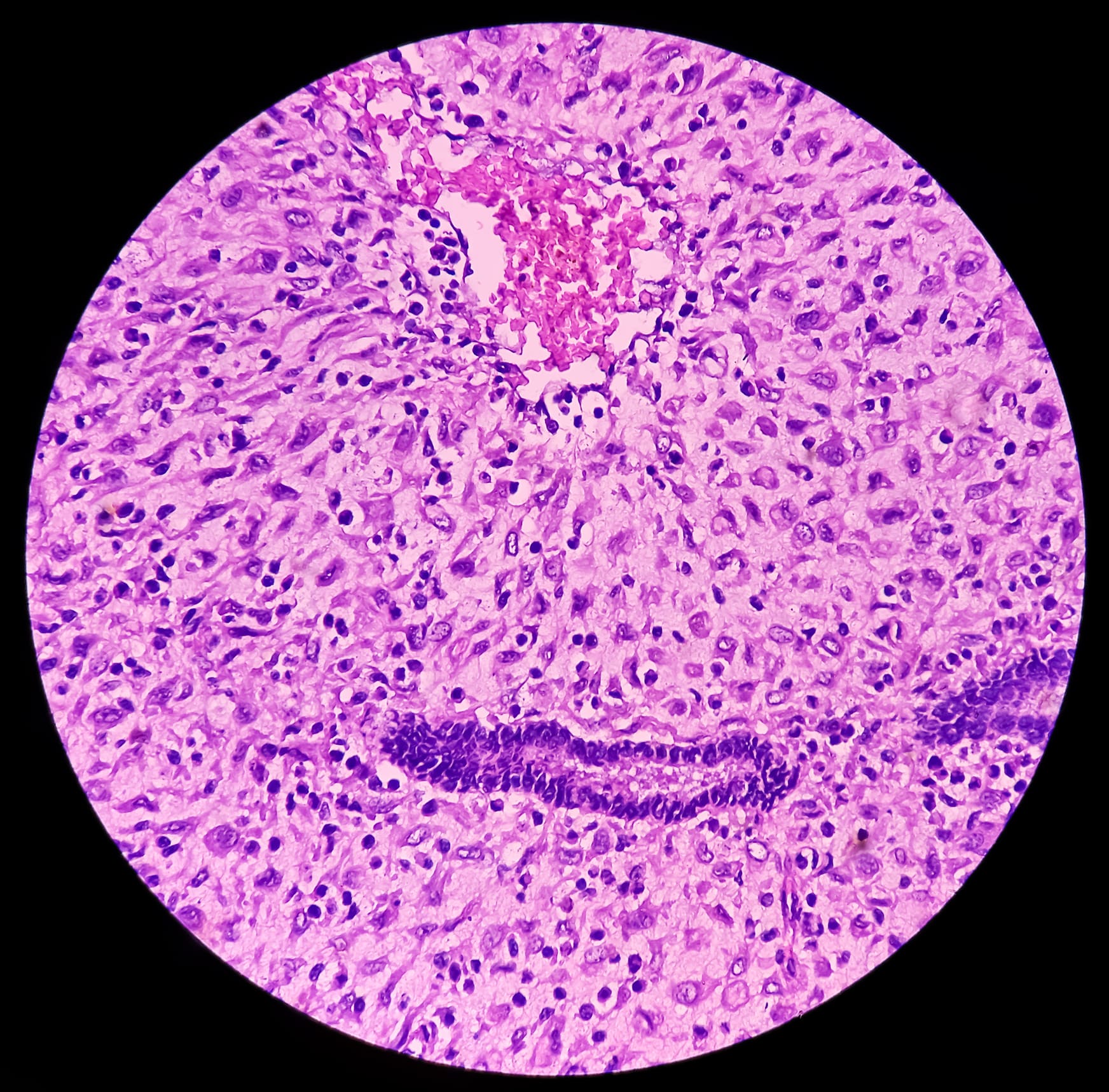
Cutaneous Rhabdomyosarcoma Often Represents Transdifferentiated Melanoma
Overview
A study of 13 patients with cutaneous epithelioid or pleomorphic rhabdomyosarcoma revealed that most tumors were actually transdifferentiated melanomas. Despite lacking conventional melanoma markers, molecular and epigenetic analyses supported melanoma lineage in the majority of cases.
Background
Cutaneous epithelioid and pleomorphic rhabdomyosarcomas are rare tumors that can be challenging to diagnose due to overlapping features with other malignancies. Melanoma is known to occasionally lose typical melanocytic markers and mimic sarcomas, complicating histopathologic classification. Accurate diagnosis is critical as it influences treatment decisions, including the potential use of immunotherapy. This study employed comprehensive molecular and epigenetic profiling to clarify the nature of these ambiguous tumors.
Data Highlights
Parameter
Findings
Patient Age Range
62-90 years (median 83)
Tumor Location
Predominantly head and neck
Immunohistochemistry
All tumors positive for desmin, myogenin, MyoD1; negative for S100 (12/12), SOX10 (10/10), Melan A (9/9), HMB45 (5/5)
DNA Methylation Profiling
9 cases tested; 7 clustered with desmoplastic melanoma
Mutational Signature
6 cases evaluable; all showed UV light signature
Tumor Mutation Burden
5 cases interpretable; 4 exceeded 10 mutations/Mb
TERT Promoter Mutations
5 of 7 sequenced cases positive
NRAS Q61R Expression
1 of 13 tumors positive
BRAF Mutations
All negative for V600E; 1 case with non-V600E BRAF p.G455R mutation
PRAME Immunohistochemistry
Positive in 3 of 8 transdifferentiated melanomas; negative in all 3 non-melanocytic tumors
Clinical Outcome
1 patient had axillary metastasis resolution after pembrolizumab, alive without disease at 25 months
Key Findings
Majority (10/13) of tumors originally diagnosed as cutaneous rhabdomyosarcoma were reclassified as transdifferentiated melanoma based on molecular and epigenetic data.
All tumors expressed skeletal muscle markers (desmin, myogenin, MyoD1) but lacked conventional melanoma markers (S100, SOX10, Melan A, HMB45).
DNA methylation profiling clustered most tumors with desmoplastic melanoma, supporting melanocytic origin despite negative immunohistochemistry.
Mutational analyses revealed ultraviolet light signatures and high tumor mutation burdens consistent with melanoma.
TERT promoter mutations were common, further indicating melanoma lineage.
PRAME immunohistochemistry showed potential diagnostic utility but with limitations.
Clinical Implications
Clinicians should consider melanoma in the differential diagnosis of poorly differentiated cutaneous tumors with rhabdomyosarcomatous features, especially in sun-damaged skin. Comprehensive molecular testing, including DNA methylation profiling, may be essential to avoid misdiagnosis and guide appropriate treatment, such as immunotherapy. Awareness of melanoma's ability to lose conventional markers and mimic sarcoma is critical for accurate diagnosis.
Conclusion
This study demonstrates that many cutaneous epithelioid or pleomorphic rhabdomyosarcomas represent transdifferentiated melanomas with distinct molecular and epigenetic profiles. Incorporating advanced molecular diagnostics can improve diagnostic accuracy and impact patient management.
Related Resources & Content
Ulici et al., Modern Pathology, 2024 -- Cutaneous Rhabdomyosarcoma May Mask Melanoma
A two-marker methylated DNA assay identified endometrial cancer with 96% sensitivity in self-collected vaginal fluid — but prospective validation is needed before it can reduce biopsies in routine care.