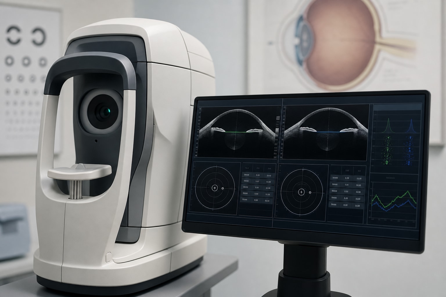
Ocular Biometric Features and Interocular Variability in Pediatric High Myopia: Insights for Early Risk Assessment
-
By
-
Siqi Zhang
-
Xi Wang
-
Zhaoxing Ding
-
Qi Zhao
-
May 25, 2026
Clinical Report: Ocular Biometric Features and Interocular Variability in Pediatric High Myopia
Overview
This study investigates the ocular biometric characteristics of pediatric high myopia, revealing significant correlations between age, axial length (AL), and axial length/corneal radius (AL/CR) ratio. The findings underscore the need for enhanced monitoring and intervention strategies for children with high myopia.
Background
The rising prevalence of myopia, particularly high myopia in children, poses significant public health challenges. Early-onset high myopia increases the risk of severe ocular complications later in life, necessitating a robust monitoring system for structural indicators beyond traditional refractive measurements. Understanding the biometric characteristics of high myopia is crucial for developing effective prevention and management strategies.
Data Highlights
| Parameter | Value |
|---|---|
| Average SE | −8.87 ± 2.58D |
| Average AL | 26.41 ± 1.33 mm |
| Average AL/CR | 3.38 ± 0.19 |
| Correlation of Age with AL | r = 0.485, P < 0.001 |
| Correlation of Age with AL/CR | r = 0.505, P < 0.001 |
| Annual AL Growth Rate | 0.224 ± 0.264 mm/year |
Key Findings
- 84 pediatric patients with high myopia were analyzed, with a mean SE of −8.87 ± 2.58D.
- Significant positive correlations were found between age and both AL (r = 0.485) and AL/CR ratio (r = 0.505).
- Age was an independent predictor of both AL and AL/CR, with a growth rate of 0.200 mm/year for AL.
- 27.7% of patients exhibited anisometropia with | ΔSE| ≥ 2.0D.
- The annual growth rate of AL in the follow-up subset was approximately 0.224 ± 0.264 mm/year.
Clinical Implications
Clinicians should prioritize monitoring axial length and AL/CR ratios in children with high myopia to assess long-term risks. Enhanced individualized monitoring and intervention strategies are essential, particularly for patients with significant binocular asymmetry.
Conclusion
The study highlights the critical need for early risk assessment and tailored management strategies in pediatric high myopia, emphasizing the importance of ocular biometric indicators in clinical practice.
Related Resources & Content
- Frontiers in Medicine, 2026 -- Age-related changes in central corneal thickness and their association with ocular biometric parameters in children
- Frontiers in Pediatrics, 2026 -- Age-related changes in lens thickness in children aged 3-17 years and its association with myopia and ocular biological parameters
- Ophthalmology Management, 2024 -- DIFFERENTIATING MYOPIA AND GLAUCOMA
- IMI—2025 Digest - PMC
- Frontiers in Ophthalmology — Diagnostic Burdens in High Myopia
- Spherical Equivalent Refraction versus Axial Length for Monitoring Childhood Myopia
- Axial length normative data and percentile curves for schoolchildren in Taiwan
- Orthokeratology, 0.04% Atropine, and 0.01% Atropine for Myopia Control
- IMI—2025 Digest - PMC
- Axial Growth and Myopia Progression After Discontinuing Soft Multifocal Contact Lens Wear
- Five-Year Clinical Trial of the Low-Concentration Atropine for Myopia Progression (LAMP) Study: Phase 4 Report - PubMed
- Axial length/corneal radius ratio for the diagnosis of myopia in children and adolescents: a meta-analysis: Clinical and Experimental Optometry: Vol 0, No 0
- Interaction effect of age and axial length-to-corneal radius ratio on myopia in children and adolescents: a cross-sectional study in China | Journal of Translational Medicine | Springer Nature Link
- Short-Term Axial Length Changes Predict Progression of Myopic Maculopathy in Pediatric High Myopia - ScienceDirect
- Association of choroidal thickness and blood flow features with asymmetric axial lengths in children with unilateral myopic anisometropia | BMC Ophthalmology | Springer Nature Link
- Axial length and corneal curvature of normal eyes in the first decade of life | Department of Ophthalmology
Based on findings from:
Interocular asymmetry and ocular biometric patterns in pediatric high myopia: implications for early risk stratification
Siqi Zhang, Xi Wang, Zhaoxing Ding, Qi Zhao. Frontiers In Medicine, 2026.
https://www.frontiersin.org/journals/medicine/articles/10.3389/fmed.2026.1841384/full
This content is an AI-generated, fully rewritten summary based on a published scholarly article. It does not reproduce the original text and is not a substitute for the original publication. Readers are encouraged to consult the source for full context, data, and methodology.