Clinical Report: Color-Coded OCT RNFL Analysis in Glaucoma Diagnosis
Overview
Expand on the specific risks associated with overreliance on color-coded OCT RNFL maps.
Background
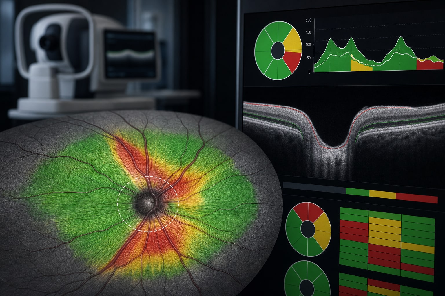
Optical coherence tomography (OCT) is a critical tool in glaucoma diagnosis and management, providing color-coded RNFL maps that are widely used in clinical practice. However, the normative data underlying these color codes often lack diversity, potentially leading to misclassification of healthy eyes and oversight of early glaucomatous changes. Understanding the limitations of these datasets is essential for accurate diagnosis and treatment.
Data Highlights
No numerical data provided in the article.
Key Findings
Color-coded OCT RNFL maps are based on limited normative datasets that may not represent diverse populations.
Mean RNFL thickness varies significantly with ancestry, axial length, and optic disc size.
Misclassification can occur in patients with high myopia or those with large optic discs, leading to false positives or negatives.
Clinicians should interpret OCT results as statistical summaries rather than definitive diagnoses.
Greater transparency in OCT reporting could improve clinical decision-making.
Clinical Implications
Clinicians should be cautious when interpreting OCT results, particularly in populations that diverge from the normative datasets. A comprehensive assessment that includes visual fields, optic disc evaluation, and patient history is crucial for accurate glaucoma diagnosis.
Conclusion
The reliance on color-coded OCT outputs in glaucoma diagnosis poses significant risks due to the limitations of the underlying normative data. A more holistic approach to patient assessment is necessary to ensure accurate diagnosis and effective management.