Clinical Report: CT-derived Model for Predicting Endotracheal Tube Dimensions in Infants
Overview
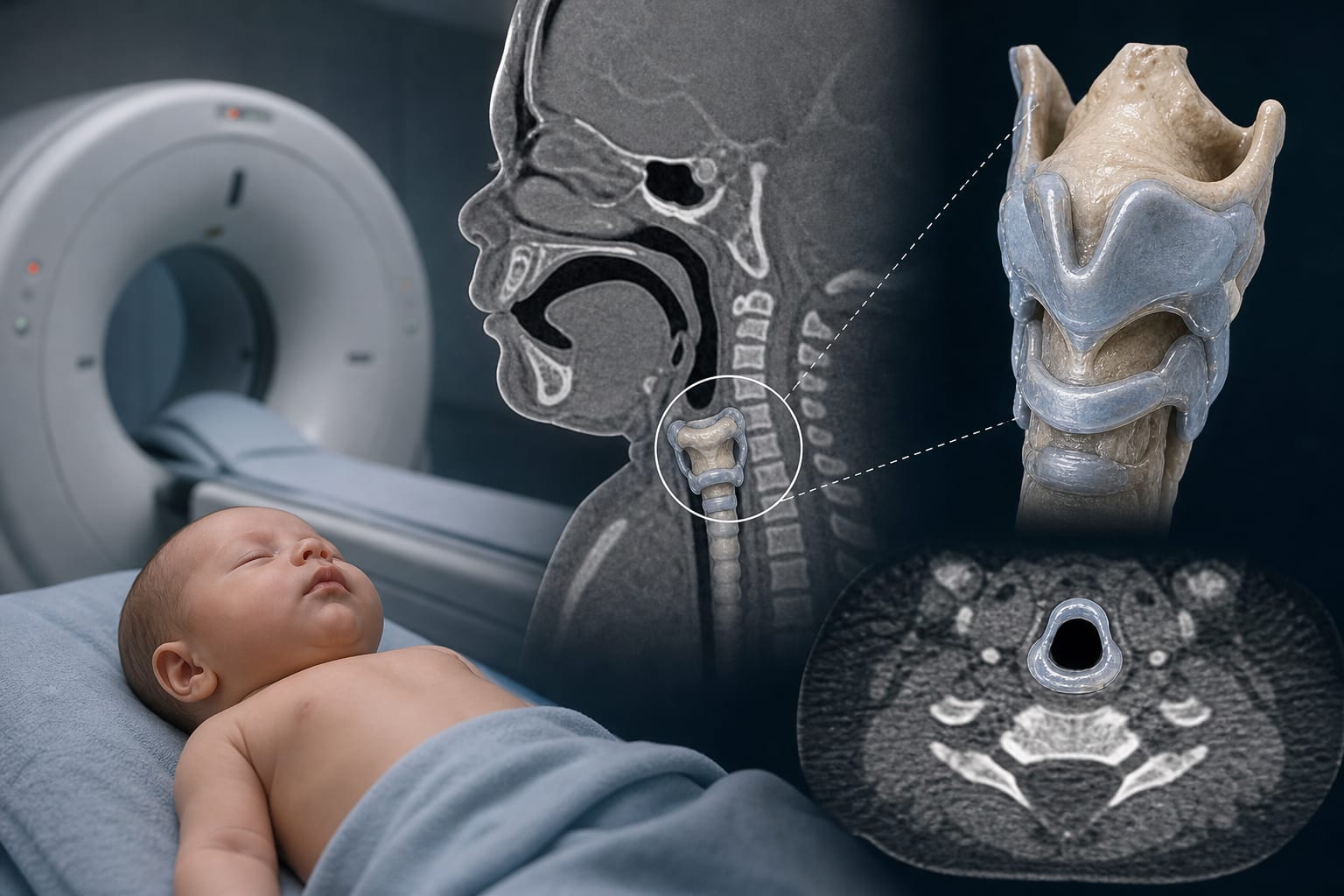
This study developed a CT-derived model to predict endotracheal tube sizes in infants under one year, emphasizing the influence of age and prematurity on cricoid cartilage dimensions. The findings suggest that individualized airway management could improve clinical outcomes in this vulnerable population.
Background
Accurate endotracheal tube (ETT) sizing is critical in infants due to their unique airway anatomy, which differs significantly from older children and adults. Traditional methods for estimating ETT size may not be reliable, particularly for infants under one year and preterm infants. This study addresses the need for more precise tools to guide airway management in this high-risk group.
Data Highlights
Parameter
Formula
Cricoid cartilage diameter (full-term)
6.119 + 0.005 × age
Cricoid cartilage diameter (preterm)
6.119 + 0.005 × age - 1.261
Cricoid-to-carina distance
35.725 + 0.044 × age
Key Findings
Age and preterm birth are independent factors affecting cricoid cartilage diameter.
The shortest diameter of cricoid cartilage can be predicted using specific formulas based on age.
The distance from cricoid cartilage to carina is significantly influenced by age.
Preterm infants may require smaller ETTs than full-term infants of the same age.
CT imaging provides a direct measurement of airway dimensions, enhancing prediction accuracy.
Clinical Implications
Clinicians should consider age and prematurity when selecting ETT sizes for infants under one year to minimize complications. The developed CT-derived formulas can aid in individualized airway management, potentially improving patient outcomes during intubation.
Conclusion
The study highlights the importance of individualized airway management in infants, with CT-derived models offering a promising approach for accurate ETT sizing. Further validation is necessary to generalize these findings to broader clinical practice.