High-frequency ultrasound features of cutaneous hidrocystoma: a case series with imaging-pathology correlation - Report - MDSpire

High-frequency ultrasound features of cutaneous hidrocystoma: a case series with imaging-pathology correlation

  • By

  • Lina Kong

  • Xiao Tang

  • Kunming Pu

  • Xiachuan Qin

  • June 26, 2026

  • 0 min

Share

Clinical Report: Ultrasound Characteristics of Cutaneous Hidrocystoma

Overview

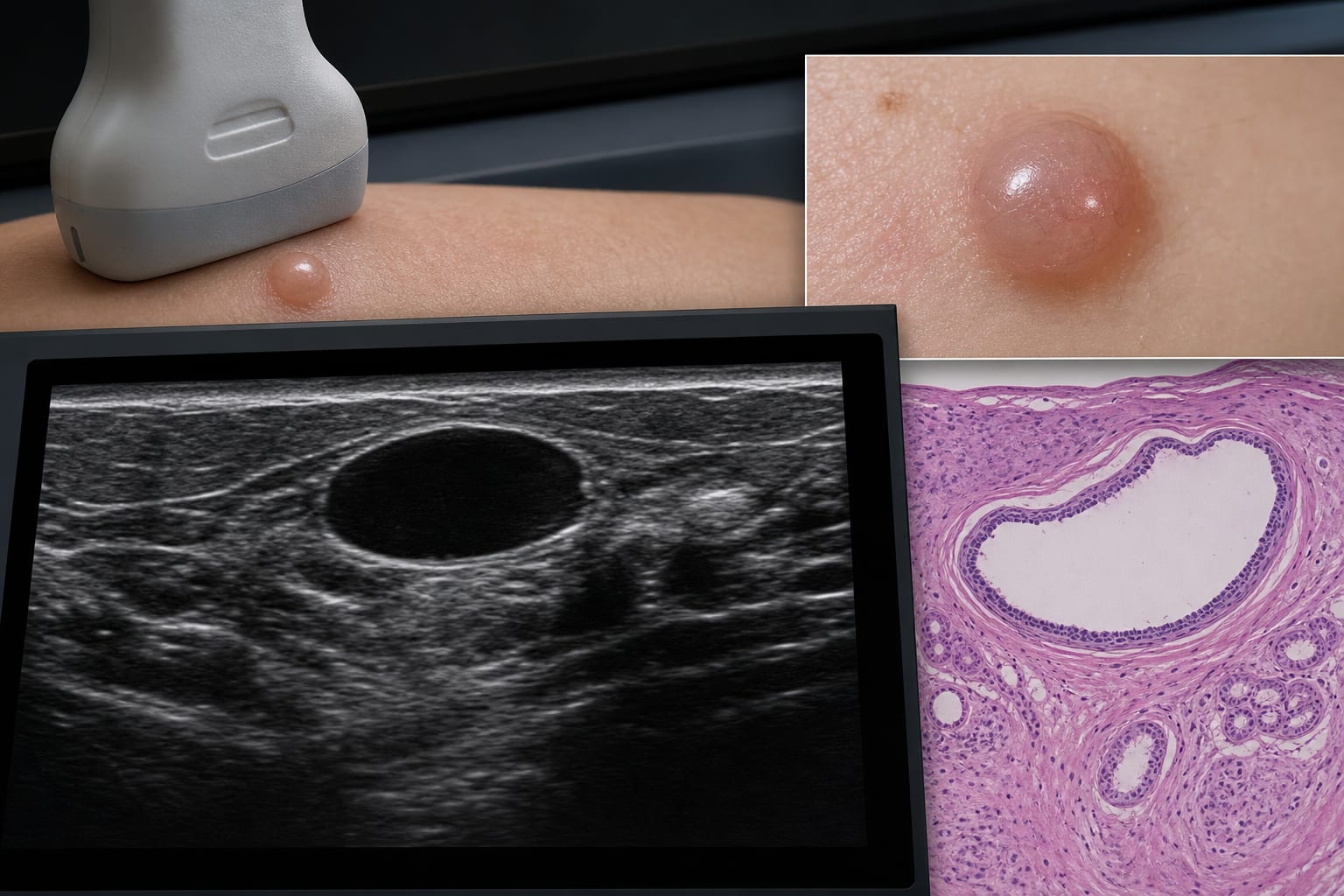
This study describes the ultrasound features of cutaneous hidrocystoma and correlates these findings with histopathology. High-frequency ultrasound (HFUS) provides information for lesion characterization, although definitive preoperative diagnosis remains challenging due to overlapping features with other lesions.

Background

Cutaneous hidrocystoma is a benign cystic lesion of sweat gland origin that poses diagnostic challenges due to its nonspecific clinical presentation. Accurate preoperative diagnosis is crucial for effective surgical planning, and while clinical examination and dermoscopy are standard, HFUS may provide additional information for lesion characterization. This study aims to systematically evaluate HFUS features in hidrocystoma and their correlation with histopathologic findings.

Data Highlights

CharacteristicFindings
Number of lesions11
LocationHead and neck region (8 lesions)
SizeMost < 1.5 cm (10/11, 90.9%)
EchogenicityHeterogeneous (36.4%), Anechoic (27.3%), Hypoechoic (18.2%), Hypoechoic with projections (18.2%)
VascularityDetected in 2 lesions (18.2%)

Key Findings

  • Eleven patients with pathologically confirmed hidrocystoma were analyzed.
  • Lesions predominantly located in the head and neck region.
  • Most lesions measured less than 1.5 cm in diameter.
  • Sonographic appearances included heterogeneous echogenicity and anechoic features.
  • Preoperative ultrasound interpretations did not identify any lesions as hidrocystoma.

Clinical Implications

HFUS may assist in localizing cutaneous hidrocystoma and narrowing the differential diagnosis, but the overlap in ultrasound features with other lesions may limit its utility for definitive preoperative diagnosis.

Conclusion

HFUS provides insights into the characteristics of cutaneous hidrocystoma, aiding in surgical planning despite challenges in achieving a definitive preoperative diagnosis.

Related Resources & Content

  1. Frontiers in Medicine, 2026 -- High-Frequency Ultrasound Features of Cutaneous Hidrocystoma: A Case Series with Imaging-Pathology Correlation
  2. Dermatology and Therapy — Beyond Drainage: Clinical–Ultrasound Correlation Supports a Broader Definition of the Active Tunnel in Hidradenitis Suppurativa
  3. Frontiers in Ophthalmology — Poroid hidradenoma of the eyelid: a case report
  4. Frontiers in Medicine — High-frequency ultrasound combined with superb microvascular imaging for differential diagnosis of basal cell carcinoma and seborrheic keratosis: a retrospective analysis with nomogram implementation
  5. Diagnostic Imaging Techniques for Evaluating Renal Tumors and Assessing Treatment Response
  6. https://efsumb.org/wp-content/uploads/2020/12/ECB2nd-ch13-DERMATOLOGY_rv2026.pdf
  7. S1 guideline for imaging diagnostics for skin diseases - Deußing - 2025 - JDDG: Journal der Deutschen Dermatologischen Gesellschaft - Wiley Online Library
  8. WHO classification of skin tumours: key updates in the fifth edition - PMC
  9. Frontiers | High-Frequency Ultrasound Features of Cutaneous Hidrocystoma: A Case Series with Imaging-Pathology Correlation
  10. Very-High-Frequency Ultrasonographic Profiling Characteristics of Nodular Hidradenoma: A Retrospective Analysis - PubMed
  11. Dermoscopy-guided high-frequency ultrasound: Principles and applications in dermatology - PubMed
  12. Value of Diagnosis and Management for Skin Lesions by Integration of High-Frequency Ultrasound in Daily Practices: A Prospective Study - PubMed
  13. AIUM Practice Parameter for Documentation of an Ultrasound Examination, 2025 Revision - PubMed

Original Source(s)

Related Content