Differential infiltration of CD4+ and CD8+ T cells and expression of PD-L1 in paired biopsy and resection specimens of gastric and colorectal adenocarcinomas - Report - MDSpire
Advertisement
Differential infiltration of CD4+ and CD8+ T cells and expression of PD-L1 in paired biopsy and resection specimens of gastric and colorectal adenocarcinomas
Comparative Analysis of CD4+ and CD8+ T Cell Infiltration and PD-L1 Expression
Overview
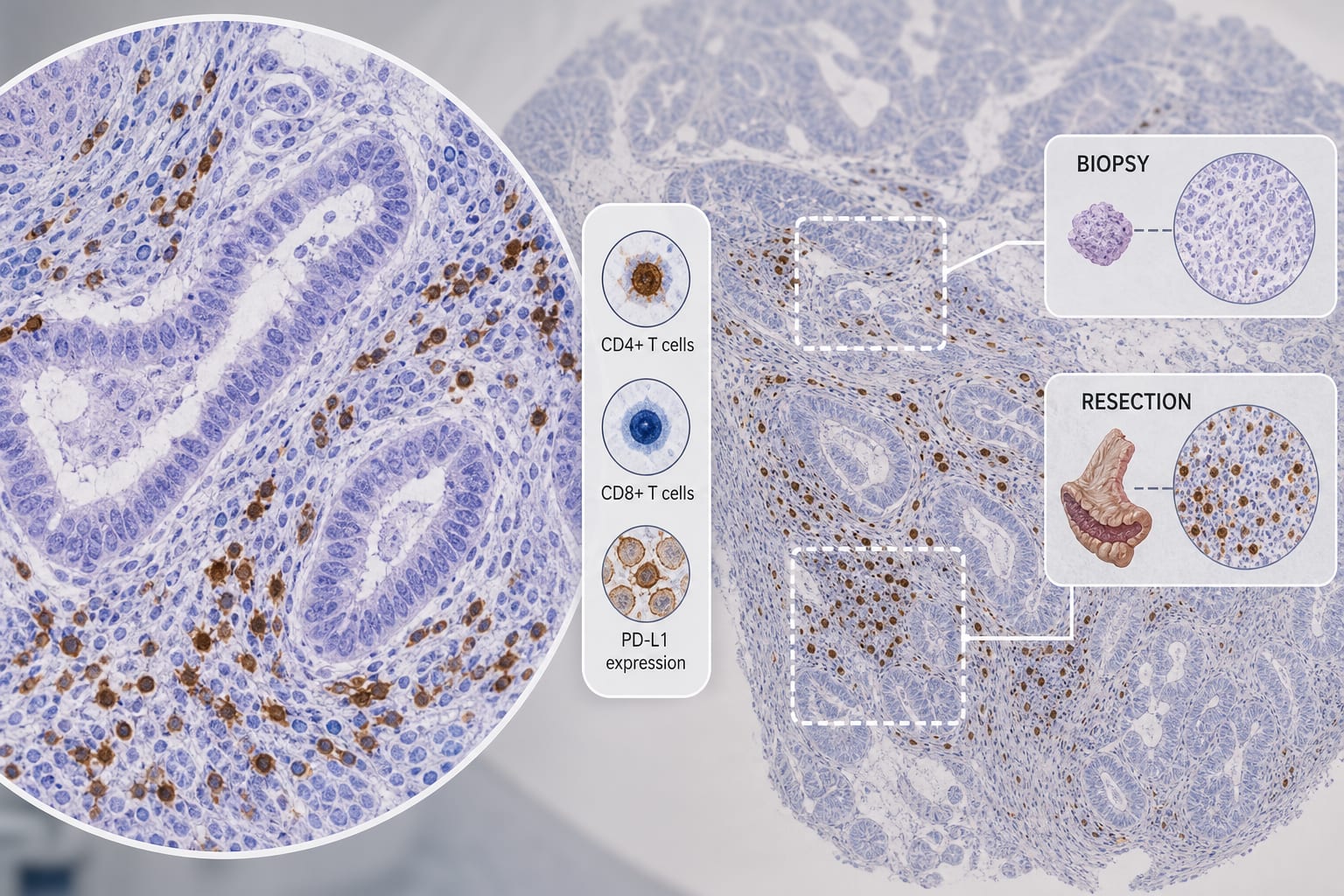
This study compares CD4+ and CD8+ T cell infiltration and PD-L1 expression in biopsy versus resection specimens of gastric and colorectal adenocarcinomas. Findings indicate that resection specimens exhibit significantly higher immune cell densities and PD-L1 expression.
Background
Understanding the immune microenvironment in gastric and colorectal adenocarcinomas is crucial. Biopsy specimens are often used for initial assessments, but their reliability in representing the tumor's immune landscape is uncertain.
Data Highlights
Finding
Value
CD4+ T cell density in gastric adenocarcinoma (resection vs biopsy)
Higher in resection (P<0.05)
CD8+ T cell density in colorectal adenocarcinoma (resection vs biopsy)
Higher in resection (P=0.0446)
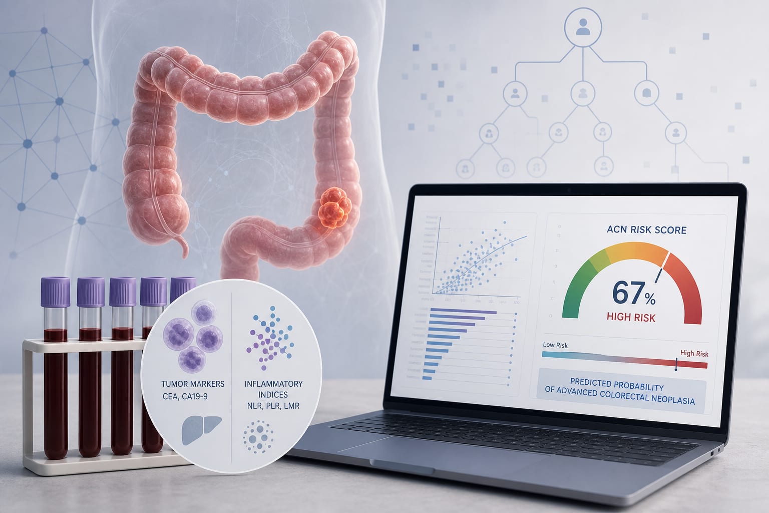
Correlation of CD4+ T cells with CA19-9 in gastric cancer
R=0.523, P=0.026
Correlation of CD4+ T cells with Ki-67 in colorectal cancer
R=-0.370, P=0.019
Preoperative neutrophil-to-lymphocyte ratio correlation with tumor diameter