Changes in retinal venous diameter in proliferative diabetic retinopathy patients with diabetic macular edema following faricimab treatment: an observational study - Report - MDSpire
Advertisement
Changes in retinal venous diameter in proliferative diabetic retinopathy patients with diabetic macular edema following faricimab treatment: an observational study
Clinical Report: Assessment of Retinal Venous Diameter in PDR Patients
Overview
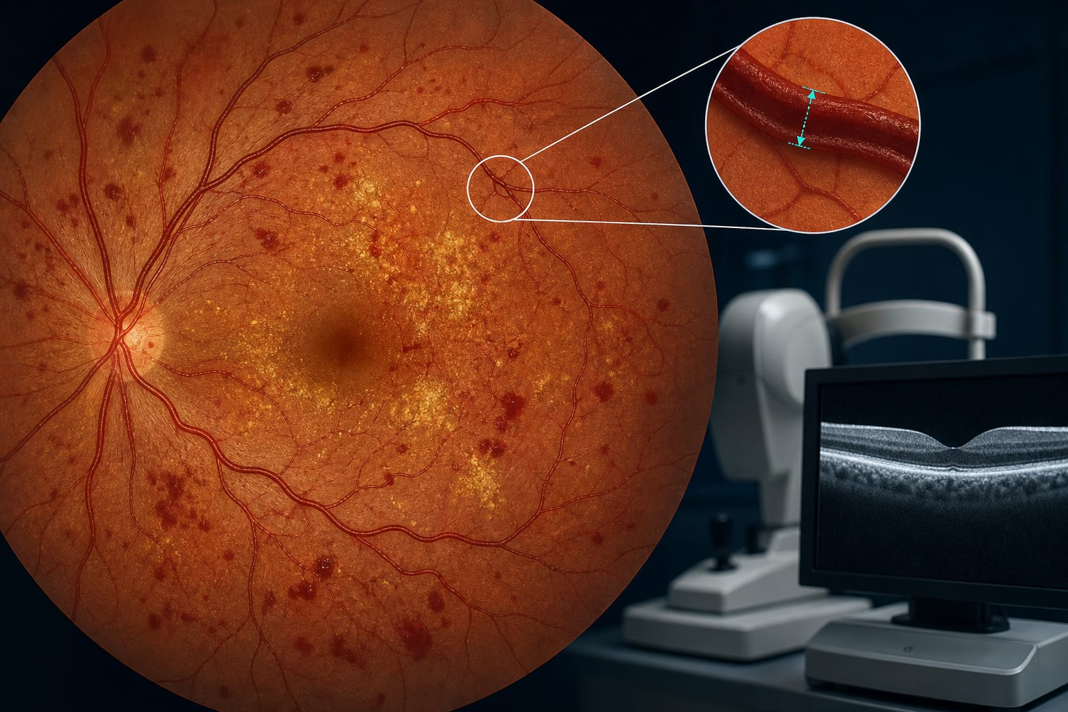
This study evaluates the impact of intravitreal faricimab on retinal venous diameter in patients with proliferative diabetic retinopathy and diabetic macular edema. Significant improvements in best-corrected visual acuity and central foveal thickness were observed, alongside a notable reduction in retinal venous diameter at 6 months post-treatment.
Background
Diabetic retinopathy is a leading cause of blindness among the working-age population, with diabetic macular edema being a critical manifestation that can lead to irreversible vision loss. Intravitreal anti-VEGF therapy is a first-line treatment for diabetic macular edema, and faricimab represents a novel approach by targeting both VEGF-A and angiopoietin-2. Understanding the effects of such therapies on retinal morphology is essential for optimizing treatment strategies.
Data Highlights
Parameter
Baseline
1 Month
3 Months
6 Months
BCVA (logMAR)
0.66 ± 0.18
0.52 ± 0.16
0.48 ± 0.18
0.40 ± 0.15
CFT (μm)
472.34 ± 47.23
381.35 ± 40.75
327.30 ± 43.40
297.56 ± 35.81
MA Count
-
-
72.71 ± 20.53
63.06 ± 17.08
HE Area
-
-
-
14 patients showed significant reduction
NV Area (mm²)
0.98 ± 0.17
0.17 ± 0.10
0.17 ± 0.09
0.17 ± 0.08
Retinal Venous Diameter (μm)
-
-
-
299.40 ± 19.56
Key Findings
Significant improvement in BCVA at 1, 3, and 6 months post-treatment (P < 0.05).
CFT significantly reduced at all time points compared to baseline (P < 0.05).
MA count decreased significantly at 3 and 6 months (P < 0.05).
NV area significantly reduced at all follow-up points (P < 0.05).
Retinal venous diameter showed a significant reduction at 6 months (P < 0.05).
Clinical Implications
The findings suggest that intravitreal faricimab may contribute to improvements in visual acuity and retinal structure in PDR patients with DME. Monitoring retinal venous diameter could provide insights into the vascular effects of treatment.
Conclusion
Intravitreal faricimab demonstrates efficacy in improving visual and anatomical outcomes in PDR patients with DME, with a notable reduction in retinal venous diameter at 6 months.