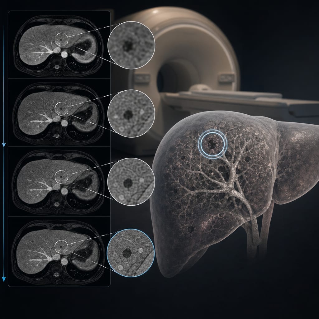
Validation of T1ρ Mapping for Hepatic Fibrosis Assessment in Chronic Liver Disease
Overview
This study clinically validates T1ρ mapping as a non-invasive MRI technique for evaluating hepatic fibrosis in patients with chronic liver disease, including those with steatotic liver disease. T1ρ mapping demonstrated promising diagnostic performance compared to established methods such as T1, ECV, T2 mapping, and MR elastography (MRE).
Background
Hepatic fibrosis is a key prognostic factor in chronic liver disease (CLD), with risk increasing alongside fibrosis stage, especially when inflammation and hepatocellular injury coexist. Non-invasive assessment methods, including serological markers and imaging techniques like MR elastography (MRE), are critical for early diagnosis and monitoring. While MRE is accurate, it requires specialized hardware, and conventional quantitative MRI techniques such as T1 and ECV mapping have limitations in the presence of hepatic steatosis. T1ρ mapping has emerged as a potentially more robust biomarker for hepatic fibrosis, warranting clinical validation.
Data Highlights
The study enrolled consecutive CLD patients undergoing liver MRI, acquiring T1ρ, T1, T2, ECV mapping, and MRE-based liver stiffness measurements. Quantitative maps were analyzed from multiple liver segments, with T1ρ mapping performed using a balanced steady-state free precession sequence with spin-lock times of 0, 30, and 60 ms. ECV was calculated from pre- and post-contrast T1 values corrected for hematocrit. MRE served as the reference standard for fibrosis staging.
Key Findings
T1ρ mapping showed strong correlation with MRE-based liver stiffness, indicating its accuracy in detecting hepatic fibrosis.
T1ρ outperformed conventional T1, ECV, and T2 mapping techniques, especially in patients with steatotic liver disease.
The technique provided robust quantitative assessment across different liver segments with good reproducibility.
T1ρ mapping does not require additional hardware beyond standard MRI scanners, facilitating broader clinical implementation.
Non-invasive T1ρ mapping may overcome limitations of serological markers and conventional MRI in fibrosis evaluation.
Clinical Implications
T1ρ mapping offers a promising non-invasive imaging biomarker for hepatic fibrosis assessment in CLD patients, including those with steatosis, potentially improving early diagnosis and monitoring without the need for invasive biopsy or specialized MRE hardware. Its integration into routine liver MRI protocols could enhance fibrosis staging accuracy and patient management.
Conclusion
T1ρ mapping is a validated, accurate, and practical MRI technique for non-invasive evaluation of hepatic fibrosis across the spectrum of chronic liver diseases, demonstrating advantages over existing quantitative MRI methods. This supports its clinical adoption for improved fibrosis assessment and monitoring.
References
Preclinical study on T1ρ as biomarker of hepatic fibrosis and portal hypertension
Meta-analyses on MRE accuracy for liver fibrosis staging
Technical descriptions of MRI sequences and quantitative mapping methods
by Narine Mesropyan, Johannes Chang, Philipp Lutz, Felix Kaiser, Oliver M. Weber, Christoph Katemann, Tatjana Dell, Dmitrij Kravchenko, Can Yueksel, Daniel Kuetting, Claus C. Pieper, Julian A. Luetkens, Alexander Isaak