Clinical Report: MRI Characteristics of Craniopharyngiomas by Age and Type
Overview
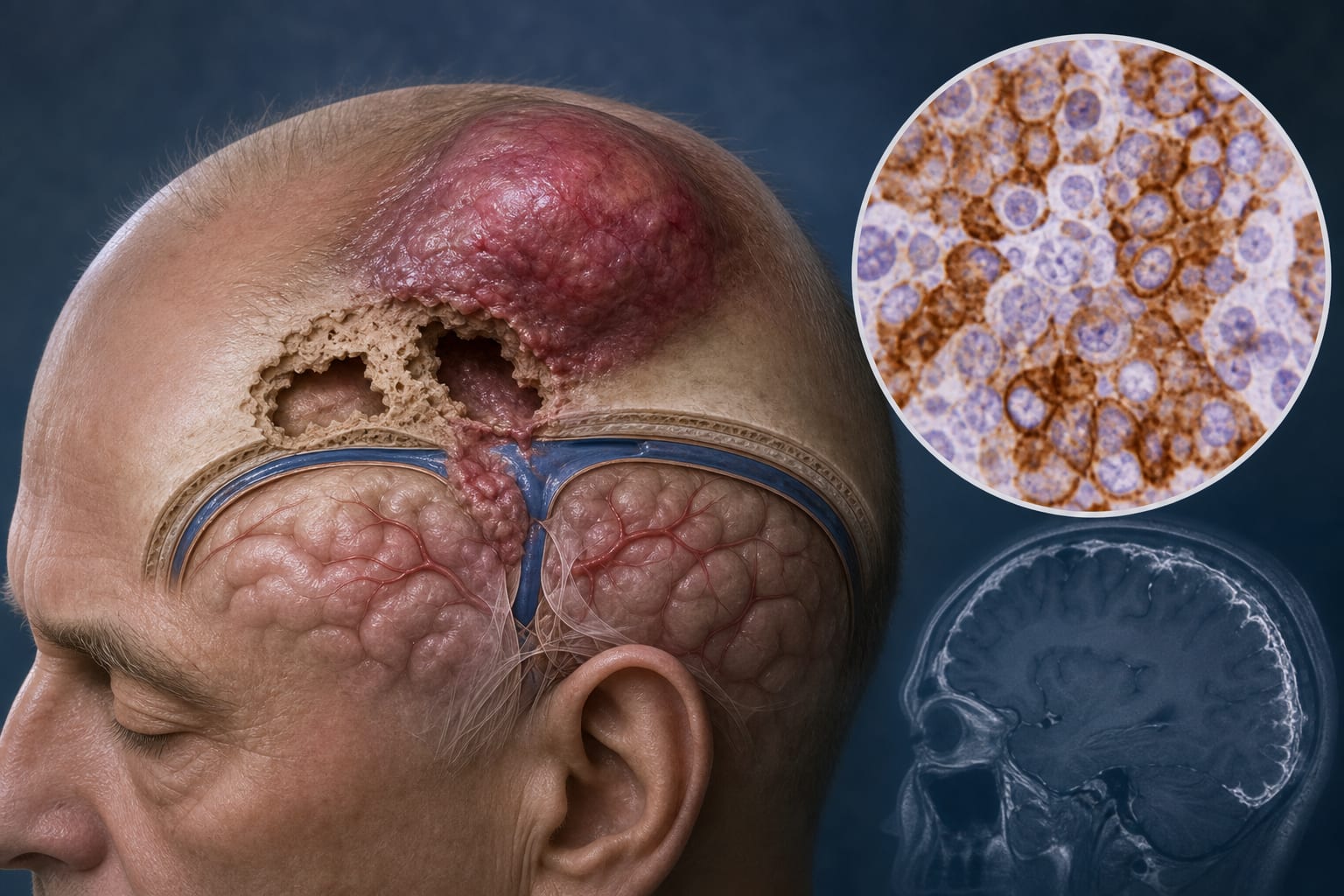
This study analyzes MRI features of craniopharyngiomas in 68 patients, revealing significant differences in tumor characteristics between juvenile and adult populations. Notably, adamantinomatous craniopharyngiomas are more prevalent in juveniles, while adults show a higher incidence of suprasellar tumors.
Background
Craniopharyngiomas are benign tumors that can lead to significant clinical complications due to their location and growth patterns. Understanding the MRI characteristics across different age groups and pathological types is crucial for accurate diagnosis and treatment planning. This study highlights the importance of age-related imaging features in guiding clinical decisions.
Data Highlights
Characteristic
Juvenile Group
Adult Group
Proportion of ACP
85.0%
56.3%
Location in Intrasellar/Suprasellar
80%
60.4%
Incidence of Calcification
55.0%
29.1%
Mean Age at Onset (ACP)
27 years
-
Mean Age at Onset (SPCP)
-
43 years
Incidence of Cystic Degeneration (ACP)
95.5%
-
Fluid-fluid Levels in ACP
20.5%
-
Key Findings
Juvenile patients have a higher prevalence of adamantinomatous craniopharyngioma (ACP) compared to adults.
Intrasellar or suprasellar locations are more common in juveniles, while adults predominantly have suprasellar tumors.
Calcification is more frequent in juvenile craniopharyngiomas.
Cystic degeneration is highly prevalent in ACP, with significant differences noted in imaging features.
Age at onset correlates with pathological subtype and MRI characteristics.
Clinical Implications
The findings suggest that MRI characteristics can aid in differentiating between craniopharyngioma subtypes based on patient age. Clinicians should consider these imaging features when planning surgical interventions and postoperative management to optimize patient outcomes.
Conclusion
This study underscores the importance of age and pathological subtype in the MRI evaluation of craniopharyngiomas, which can inform clinical decision-making and improve patient management strategies.