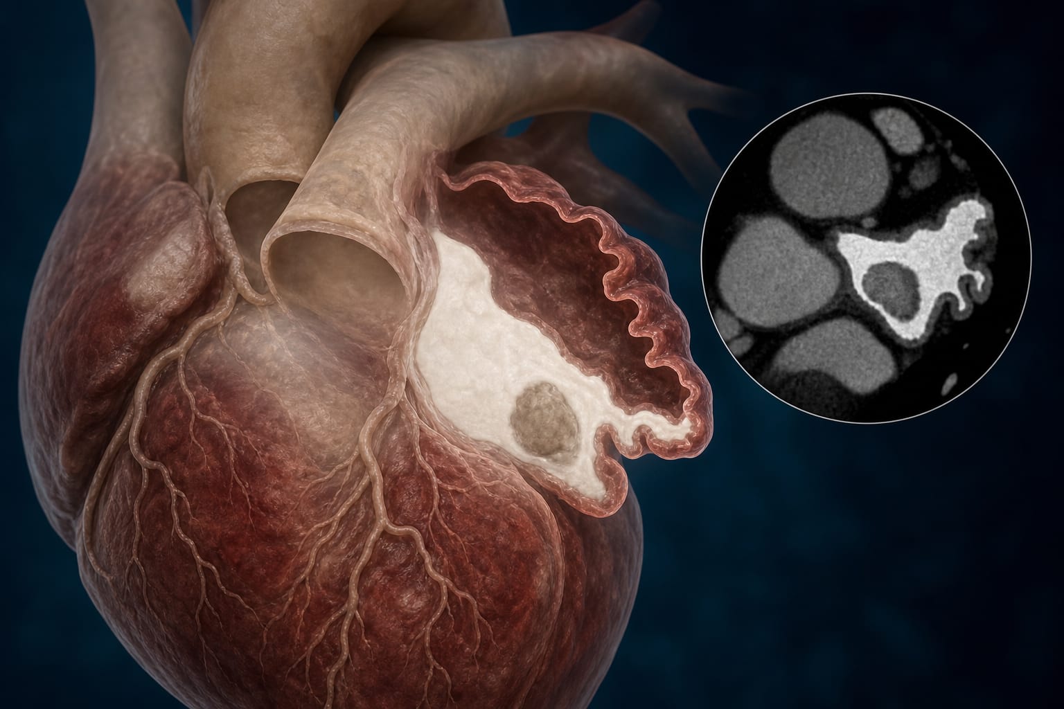
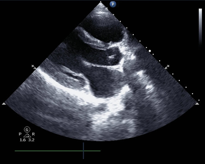
Optimizing left atrial appendage imaging: the diagnostic value of left lateral decubitus cardiac CT angiography
-
By
-
Zihao Wang
-
ChenJing Wu
-
Yi Mang
-
Zhuang Zhuang
-
Xinshi Huang
-
Zhenzhang Wang
-
June 5, 2026
-
0 min