Adenomatoid tumors of the uterus: magnetic resonance imaging features with clinical and histopathologic correlation - Report - MDSpire
Coming Soon: Introducing MDSpire News. Learn more
Conexiant’s news site is now MDSpire News. Learn more

Magnetic Resonance Imaging Characteristics of Uterine Adenomatoid Tumors: A Study of Clinical and Histopathological Correlations

  • By

  • Changhuan Chen

  • Yudian Huang

  • Ping Chen

  • Caixia Zheng

  • Chengsheng Wang

  • Mingsi Yang

  • September 8, 2026

Share

Clinical Report: Magnetic Resonance Imaging Characteristics of Uterine Adenomatoid Tumors

Overview

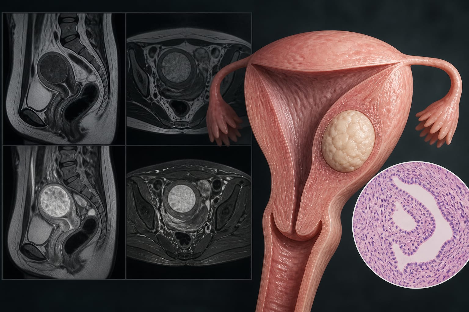
This study characterizes the MRI features of uterine adenomatoid tumors (ATs) and correlates these findings with histopathological data. The results indicate that ATs typically present as solid masses with specific imaging characteristics that can aid in preoperative diagnosis.

Background

Uterine adenomatoid tumors are rare benign neoplasms that can be misdiagnosed due to their imaging similarities with uterine leiomyomas. Accurate preoperative diagnosis is crucial to avoid unnecessary interventions and to guide appropriate treatment strategies. Limited literature exists on the MRI characteristics of these tumors, highlighting the need for comprehensive studies.

Data Highlights

CharacteristicFindings
Median Age51 years
Lesion Count17 lesions from 16 patients
Lesion Location7 intramural, 10 subserosal
Maximum Diameter9–65 mm
Lesion Composition13 solid, 2 cystic-solid, 2 cystic
Target Sign on T2WI4 lesions

Key Findings

  • Uterine ATs typically appear as solid masses without a discernible capsule.
  • Lesions may exhibit cystic degeneration or a 'target sign' on T2-weighted imaging.
  • Most lesions show mild diffusion restriction within their solid components.
  • Histopathological examination is necessary for definitive diagnosis.
  • Imaging findings correlate closely with histopathological characteristics.

Clinical Implications

Understanding the MRI characteristics of uterine ATs can improve preoperative diagnosis and reduce the risk of misdiagnosis with uterine leiomyomas. Clinicians should consider these imaging features when evaluating patients with suspected uterine masses.

Conclusion

The study provides valuable insights into the MRI characteristics of uterine adenomatoid tumors, emphasizing the importance of histopathological confirmation for accurate diagnosis.

Related Resources & Content

  1. IARC Publications Website, Female Genital Tumours, 2020 -- Adenomatoid Tumour
  2. American College of Radiology ACR Appropriateness Criteria®, Abnormal Uterine Bleeding
  3. Frontiers in Medicine — Magnetic resonance-guided focused ultrasound surgery for adenomyosis: current evidence and future directions
  4. Frontiers in Endocrinology — Identification of Diagnostic Blood Indicators Associated with Adenomyosis: A Retrospective Cohort Study
  5. European Radiology — Key Insights on MRI and CT for the Evaluation and Staging of Adnexal Masses: Recommendations from ESUR
  6. Frontiers in Medicine — Mesonephric-like adenocarcinoma of the uterine corpus: a case report
  7. Magnetic resonance-guided focused ultrasound surgery for adenomyosis: current evidence and future directions
  8. Identification of Diagnostic Blood Indicators Associated with Adenomyosis: A Retrospective Cohort Study
  9. Key Insights on MRI and CT for the Evaluation and Staging of Adnexal Masses: Recommendations from ESUR
  10. IARC Publications Website - Female Genital Tumours
  11. American College of Radiology ACR Appropriateness Criteria® Abnormal Uterine Bleeding
  12. mrms-MP-2022-0067-Takeuchi 127..135

Original Source(s)

Related Content