Clinical Report: Evaluation of Skin Anomalies Using Triple Wavelength Imaging Techniques
Background
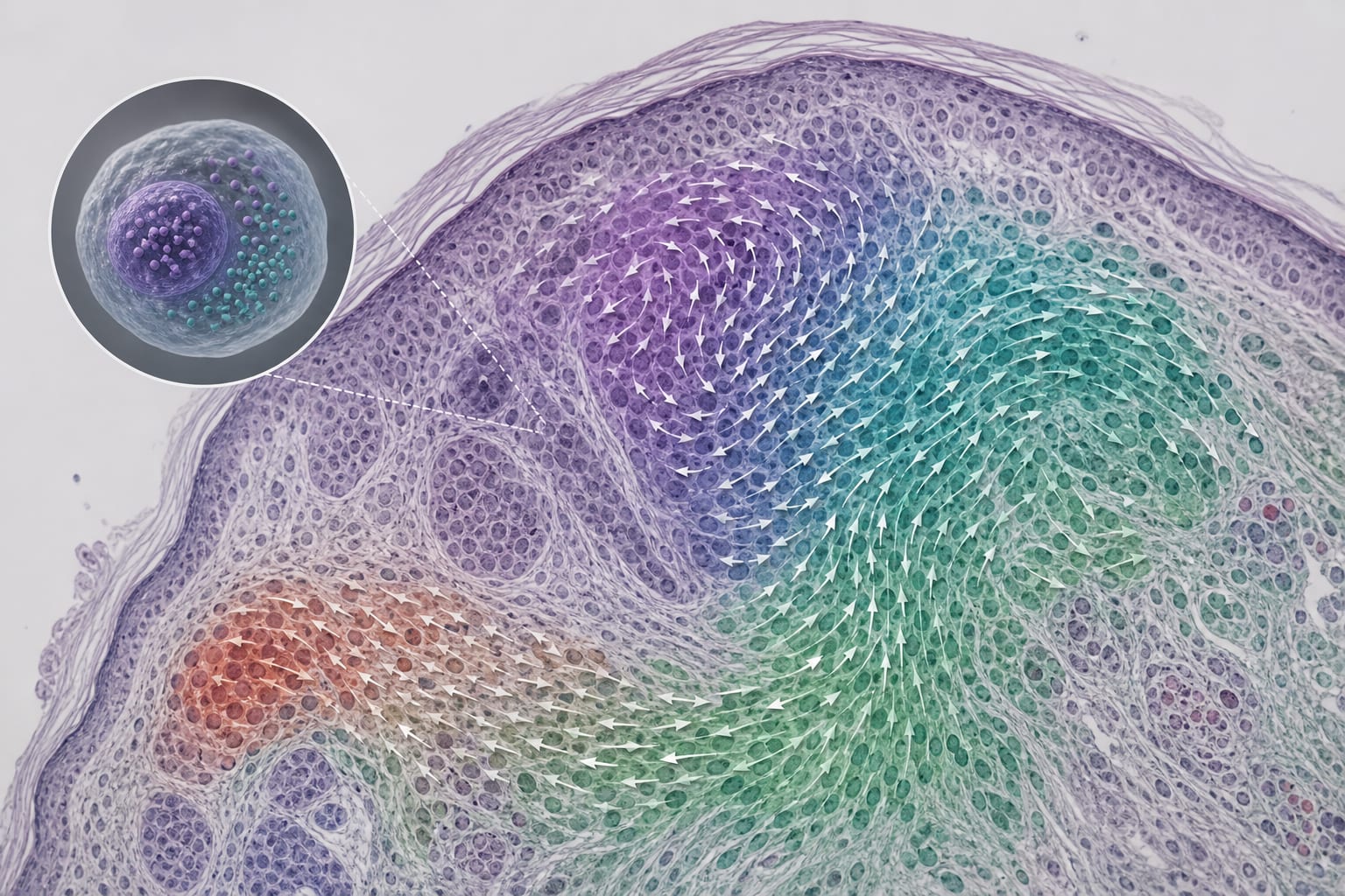
Timely detection of skin tumors, particularly malignant melanoma, is critical due to its aggressive nature and high metastatic potential. Traditional imaging methods often fall short in providing detailed spectral information necessary for accurate diagnosis.
Data Highlights
No numerical data or trial results were provided in the source material.
Key Findings
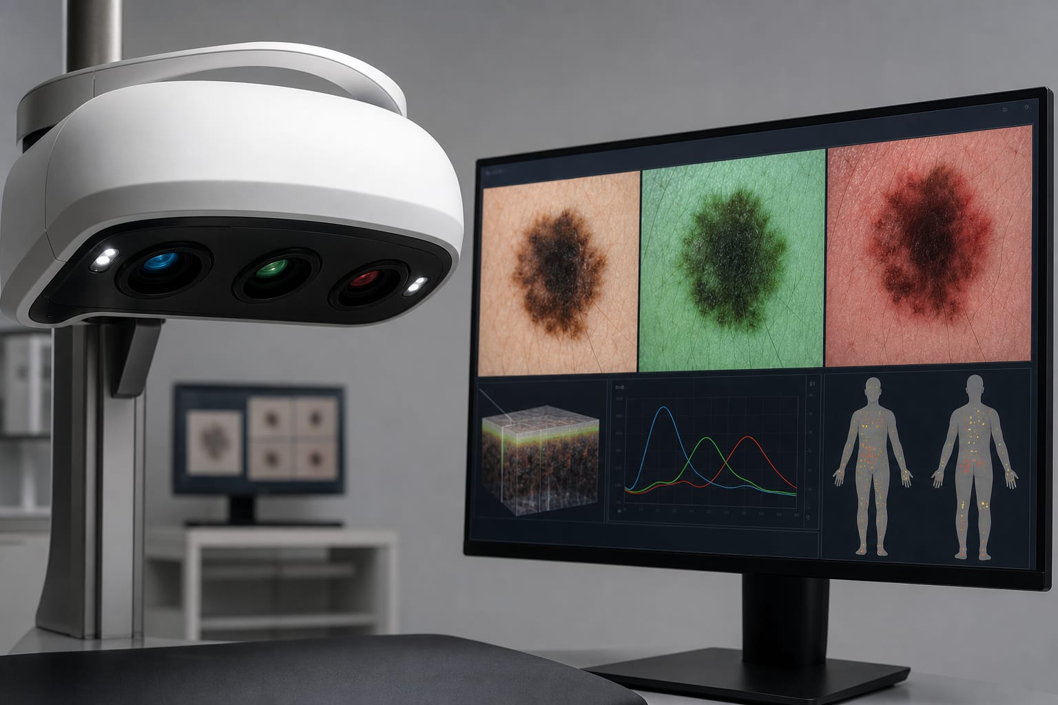
Triple wavelength imaging (3WI) allows for high-resolution spectral imaging of skin lesions.
The technique minimizes motion artifacts by capturing images in a single snapshot.
3WI has been clinically validated on volunteers with various skin malformations, including cancers.
RGB lasers are effective light sources for 3WI, ensuring uniform illumination.
The method enhances spectral selectivity with imaging bandwidths typically less than 0.1 nm.
Clinical Implications
The implementation of triple wavelength imaging could enhance diagnostic accuracy in dermatology by providing detailed spectral information.
Conclusion
Triple wavelength imaging represents a significant advancement in noninvasive skin diagnostics.