Clinical Report: Gender Variations in Resting-State EEG Spectral Power
Overview
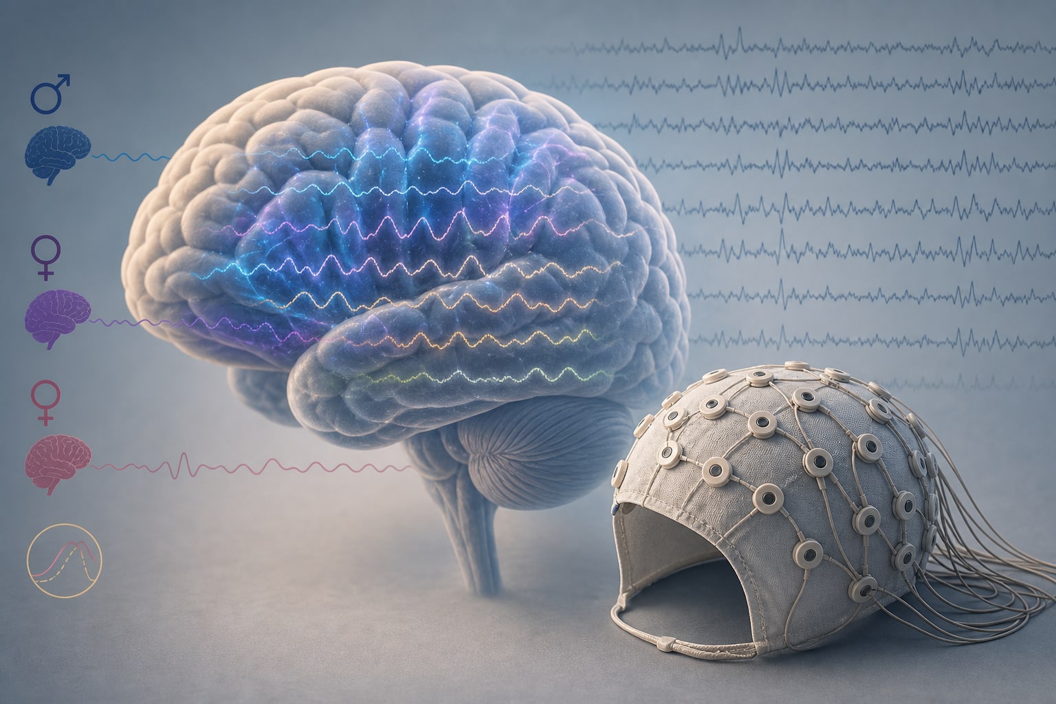
This study investigates the influence of sex hormones on resting-state EEG spectral power in healthy young adults, revealing that sex differences in spectral power are more pronounced than menstrual cycle effects.
Background
Understanding the relationship between sex hormones and brain activity is crucial for insights into cognitive and behavioral processes. Fluctuations in hormones like estradiol and progesterone across the menstrual cycle can significantly impact neural oscillations, which are measurable through EEG.
Data Highlights
No numerical data or trial data presented in the source material.
Key Findings
Sex differences in resting-state EEG spectral power were more prominent than differences between menstrual cycle phases.
No significant differences were observed between women with high versus low estradiol levels.
Widespread sex differences in high-frequency bands were noted, particularly in frontal and central-left regions.
Within females, estradiol was associated with posterior high gamma power, while progesterone correlated with theta and high beta power.
No hormone–EEG associations were significant in the male group after FDR correction.
Clinical Implications
Consideration of sex and hormonal status may be relevant in studies of brain function and EEG analysis.
Conclusion
The study highlights sex and hormonal influences on EEG spectral power.
by Angelika K. Sawicka, Aleksandra M. Zieminska, Natalia Zalewska, Adrianna Czerwińska, Katarzyna M. Michalak, Barbara Naparło, Nastaran Hamedi, Jesús S. García-Salinas, Anna B. Marcinkowska, Michal T. Kucewicz, Paweł J. Winklewski
In two population-based cohorts, metabolically unhealthy status generally showed higher dementia risk estimates, while metabolically healthy obesity was not associated with increased risk in primary analyses.