Sequential ¹⁸F‑AV45/¹⁸F‑AV1451 dual‑tracer brain PET imaging in Alzheimer's disease: amyloid‑tau deposition, diagnostic performance, cognitive associations, and modulation by APOE ε4 - Report - MDSpire
Advertisement
Sequential ¹⁸F‑AV45/¹⁸F‑AV1451 dual‑tracer brain PET imaging in Alzheimer's disease: amyloid‑tau deposition, diagnostic performance, cognitive associations, and modulation by APOE ε4
Clinical Report: Dual-Tracer PET Imaging in Alzheimer's Disease
Overview
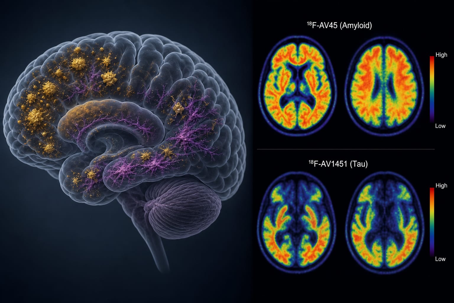
This study evaluates the efficacy of dual-tracer PET imaging using ¹⁸F‑AV45 and ¹⁸F‑AV1451 in characterizing amyloid and tau deposition in Alzheimer's disease. APOE ε4 status influences both amyloid and tau levels.
Background
Alzheimer's disease is characterized by the presence of amyloid-β plaques and tau neurofibrillary tangles, which are critical for diagnosis and understanding disease progression. The AT(N) framework emphasizes the need for in vivo imaging of these pathologies to improve diagnostic accuracy and therapeutic monitoring. Despite the importance of dual-pathology imaging, large-scale clinical data on its effectiveness remain limited.
Data Highlights
Group
Aβ SUVR
Tau SUVR
AD
Higher
Higher
MCI
Lower
Lower
HC
Lowest
Lowest
Key Findings
Whole-brain and regional SUVRs for both tracers were significantly higher in AD compared to MCI and HC (P<0.001).
The dual-tracer strategy yielded an AUC of 0.97 for distinguishing AD from HC and 0.93 for AD from MCI.
Tau deposition showed stronger cognitive correlations than Aβ deposition.
APOE ε4 carriers exhibited significantly higher Aβ and tau deposition.
Aβ and tau SUVRs were strongly correlated (r=0.65–0.81), particularly in the precuneus and inferior temporal gyrus.
Clinical Implications
The findings support the use of dual-tracer PET imaging as a tool for evaluating Alzheimer's disease pathology.
Conclusion
Dual-tracer PET imaging provides a comprehensive assessment of Alzheimer's disease-related pathology.