Endoscopic Debridement for Calcific Tendinitis at the Deep Insertion of the Gluteus Maximus
Overview
This case study presents a 53-year-old woman with calcific tendinitis at the deep insertion of the gluteus maximus, diagnosed through MRI and CT after inconclusive radiographs. Endoscopic debridement under C-arm guidance resulted in complete removal of the calcific deposit.
Background
Calcific tendinitis is commonly associated with the rotator cuff but can also affect the deep insertion of the gluteus maximus, which is less frequently reported. Accurate diagnosis is critical, as deep-seated lesions may not be visible on standard radiographs, leading to delays in treatment. Imaging techniques such as MRI and CT are essential for proper localization and management of these conditions.
Data Highlights
This case study does not contain numerical data or trial data suitable for tabulation.
Key Findings
A 53-year-old woman presented with lateral hip pain and limited range of motion unresponsive to NSAIDs.
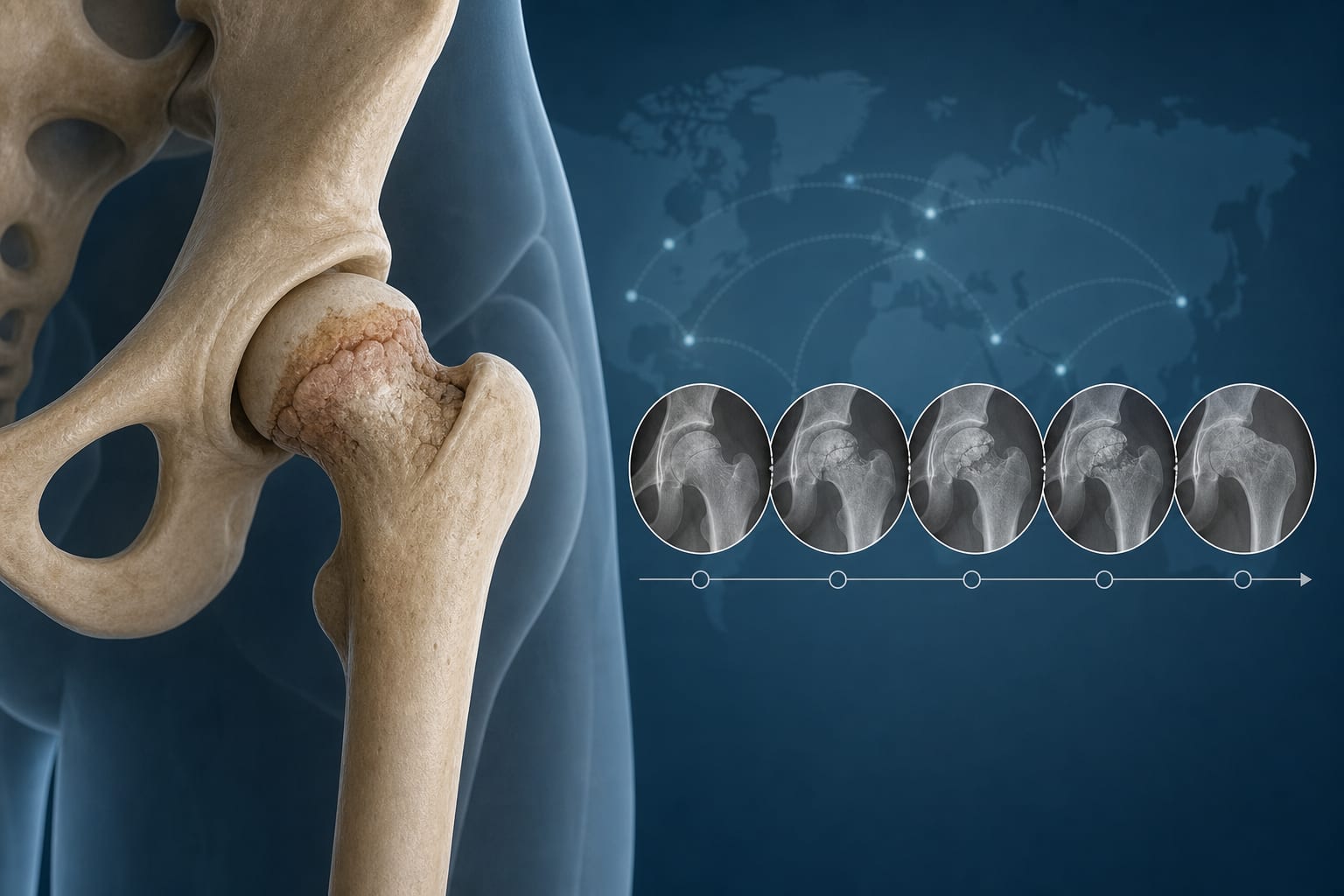
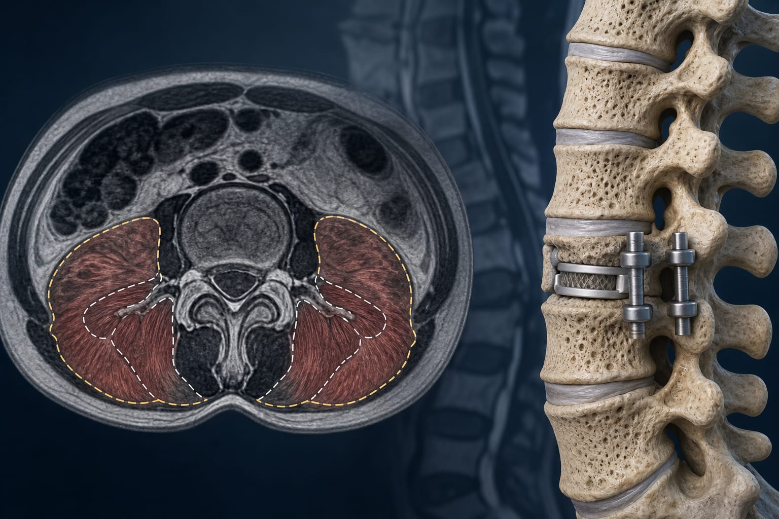
Plain radiographs were inconclusive, while MRI and CT identified inflammatory edema and a calcific deposit.
Endoscopic debridement under C-arm guidance successfully removed the calcific material.