Reliable and consistent mapping of population receptive fields in retinal disorders
Overview
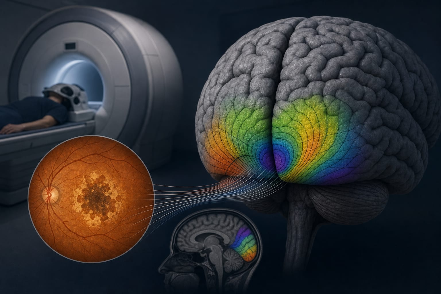
This study evaluates the reproducibility of population receptive field (pRF) mapping using 7 Tesla fMRI in patients with Stargardt disease and geographic atrophy. It highlights the potential of pRF mapping as an objective tool for assessing visual field representation in retinal disorders.
Background
Retinal disorders significantly impact visual function, and accurate assessment methods are crucial for diagnosis and treatment planning. Traditional subjective methods, such as perimetry, can be influenced by various factors, leading to variability in results. The advent of fMRI provides an objective means to assess neural responses to visual stimuli, which may enhance our understanding of visual field representation in patients with retinal diseases.
Data Highlights
No numerical data was provided in the article.
Key Findings
The study involved 22 patients: 11 with Stargardt disease and 11 with geographic atrophy.
pRF mapping at 7 Tesla demonstrated high reproducibility in healthy participants, but similar data in clinical populations was previously lacking.
Both intrasession and intersession reproducibility of pRF mapping were assessed in patients with retinal disorders.
Retinotopic mapping revealed distinct cortical representations corresponding to retinal lesions.
Higher magnetic field strengths (7 Tesla) yield improved resolution of activation maps compared to lower field strengths.
Clinical Implications
The findings suggest that pRF mapping could serve as a reliable objective tool for evaluating visual field representation in patients with retinal disorders. This may facilitate better understanding and monitoring of disease progression and treatment effects.
Conclusion
The study underscores the potential of 7 Tesla fMRI pRF mapping as a reproducible method for assessing visual field representation in patients with retinal disorders, paving the way for future research in this area.
by Maximilian Pawloff, David Linhardt, Michael Woletz, Marlene Hollaus, Georgios Mylonas, Graham E. Holder, Stefan Sacu, Christian Windischberger, Markus Ritter