A case report and literature review on imaging manifestations of immature teratoma in fetal oral cavity with concurrent intracranial abnormalities - Report - MDSpire
Advertisement
A case report and literature review on imaging manifestations of immature teratoma in fetal oral cavity with concurrent intracranial abnormalities
Clinical Report: Imaging Features of Fetal Oral Cavity Immature Teratoma
Overview
This report details the imaging characteristics of a rare case of immature teratoma in the fetal oral cavity, highlighting its association with intracranial abnormalities. The findings underscore the importance of multimodal imaging for accurate prenatal diagnosis.
Background
Fetal teratomas, particularly those located in the oral cavity, are rare and can lead to significant morbidity and mortality during the perinatal period. Accurate prenatal diagnosis is crucial for planning management and intervention strategies. This case emphasizes the need for enhanced diagnostic capabilities through advanced imaging techniques.
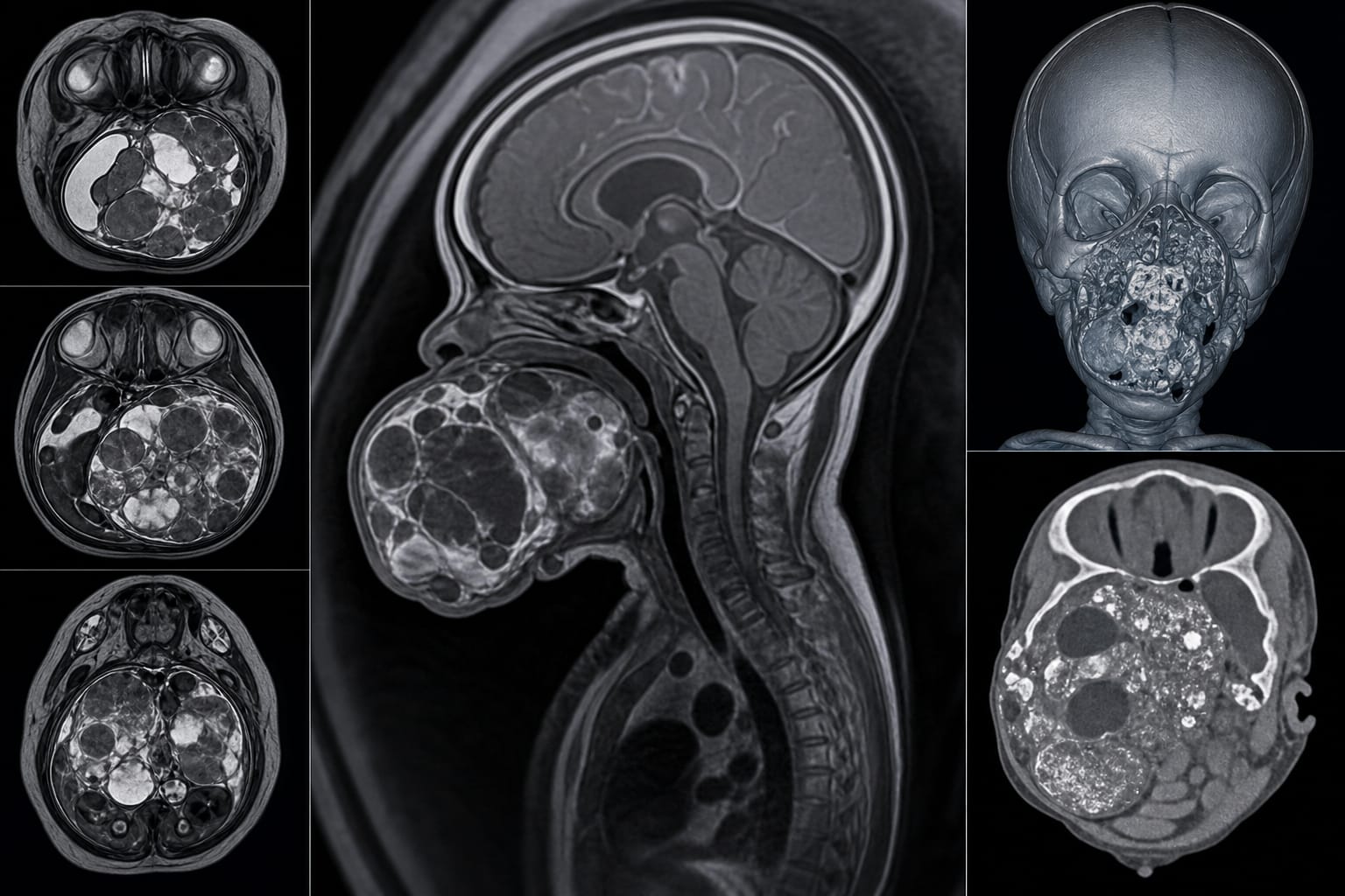
Irregular solid-cystic mass, low signal intensity on T1WI, high signal intensity on T2WI, restricted diffusion
Key Findings
Large solid-cystic mass in the fetal oropharynx associated with the maxilla and skull base.
CT imaging revealed heterogeneous density with punctate calcifications, characteristic of immature teratomas.
MRI showed low signal intensity on T1-weighted images and high signal intensity on T2-weighted images.
Pathological examination confirmed a Grade III immature teratoma with concurrent right temporal arachnoid cyst and meningocele.
Multimodal imaging aids in differentiating immature from mature teratomas based on specific imaging features.
Clinical Implications
The findings highlight the importance of using multimodal imaging techniques, such as MRI and CT, for the prenatal diagnosis of fetal teratomas. Clinicians should be aware of the potential for concurrent intracranial abnormalities, which necessitate comprehensive evaluation and planning for delivery.
Conclusion
This case study enhances understanding of the imaging features of fetal oral cavity immature teratomas and their associated complications. Improved diagnostic proficiency can lead to better management strategies for affected fetuses.