Management of a Mandibular Third Molar With Radix Paramolaris: A Case Report
-
By
-
Zak R. James, DMD
-
September 1, 2026
-
11 min
Clinical Report: Management of a Mandibular Third Molar With Radix Paramolaris
Overview
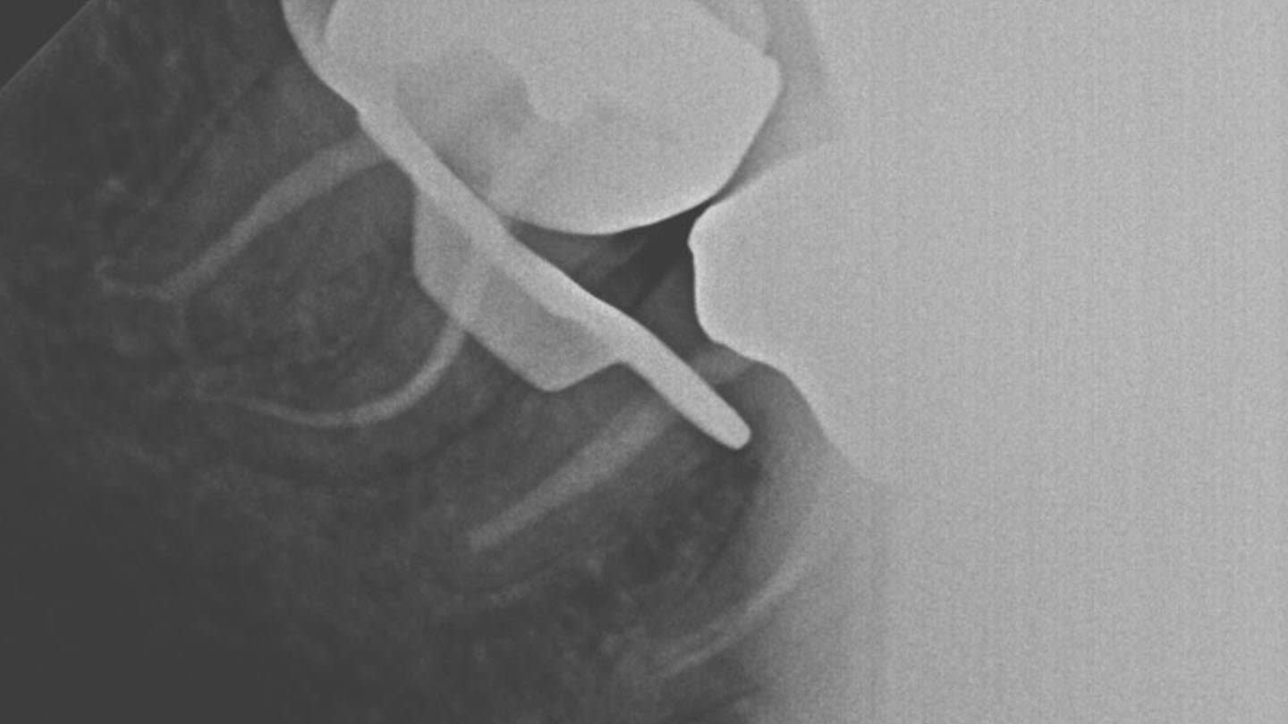
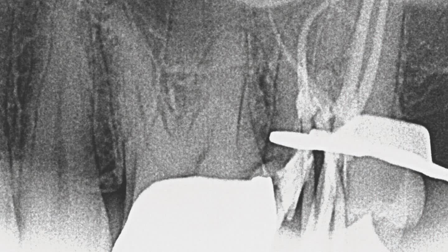
This case report details the nonsurgical root canal treatment of a mandibular third molar with a radix paramolaris in an 86-year-old patient. The treatment involved the use of cone-beam computed tomography (CBCT) to identify complex root canal anatomy, and modern instrumentation was utilized to facilitate successful management.
Background
The management of mandibular third molars can be complicated due to their complex anatomy and limited access. The presence of anatomical variations, such as radix paramolaris, can complicate treatment and increase the risk of endodontic failure if not recognized. Advanced imaging techniques, such as CBCT, are essential for effective treatment planning.
Data Highlights
No numerical or trial data provided in the article. The case highlights the importance of recognizing anatomical variations in treatment planning.
Key Findings
- The patient presented with pulpal necrosis and symptomatic apical periodontitis associated with a crowned mandibular third molar.
- CBCT imaging revealed a radix paramolaris originating from the mesiobuccal root, influencing treatment planning.
- Nonsurgical root canal treatment was performed over two appointments through a conservative access cavity to preserve the existing crown.
- Modern heat-treated nickel-titanium rotary instrumentation was utilized to manage the complex root canal anatomy.
- Failure to recognize accessory roots can lead to persistent infection and endodontic failure.
Clinical Implications
Clinicians should consider advanced imaging techniques like CBCT when faced with complex root canal anatomy in mandibular third molars.
Conclusion
This case emphasizes the importance of thorough diagnostic imaging and conservative treatment approaches in managing complex endodontic cases.
Related Resources & Content
- Giuseppe Monaco, DMD et al., Inside Dentistry, 2013 -- Coronectomy of Mandibular Third Molars
- Compendium, 2012 -- Nonsurgical Endodontic Retreatment of a Mandibular First Molar with Five Canals: 1-Year Follow-up
- Journal of the American Dental Association, 2026 -- Management of a maxillary lateral incisor with a type III palatogingival groove and 2 roots: A case report
- Compendium, 2024 -- Impacted Mandibular “Double” Third Molar: Fusion or Gemination? A Case Report
- American Dental Association, 2026 -- New ADA recommendations confirm dental imaging most effectively used in moderation
- AAE Newsroom, 2026 -- AAE and AAOMR Release Joint Position Statement on the Use of CBCT in Endodontics
- New ADA recommendations confirm dental imaging most effectively used in moderation | American Dental Association
- AAE and AAOMR Release Joint Position Statement on the Use of CBCT in Endodontics - AAE Newsroom
- The Worldwide Prevalence of Radix Molaris in Mandibular Second Molars Assessed by Cone Beam Computed Tomography: Systematic Review and Meta-analysis - PubMed
- Prevalence and anatomy of radix entomolaris and paramolaris in mandibular molars - A CBCT study at North Western India - PubMed
- Expert consensus on the management of third molar health
Based on findings from:
Management of a Mandibular Third Molar With Radix Paramolaris: A Case Report
Zak R. James, DMD. Compendium, 2026.
https://compendiumlive.com/2026/09/thirdmolar/
This content is an AI-generated, fully rewritten summary based on a published scholarly article. It does not reproduce the original text and is not a substitute for the original publication. Readers are encouraged to consult the source for full context, data, and methodology.