Clinical Report: Evaluation of Deep Learning Models Utilizing Multimodal Ultrasound
Overview
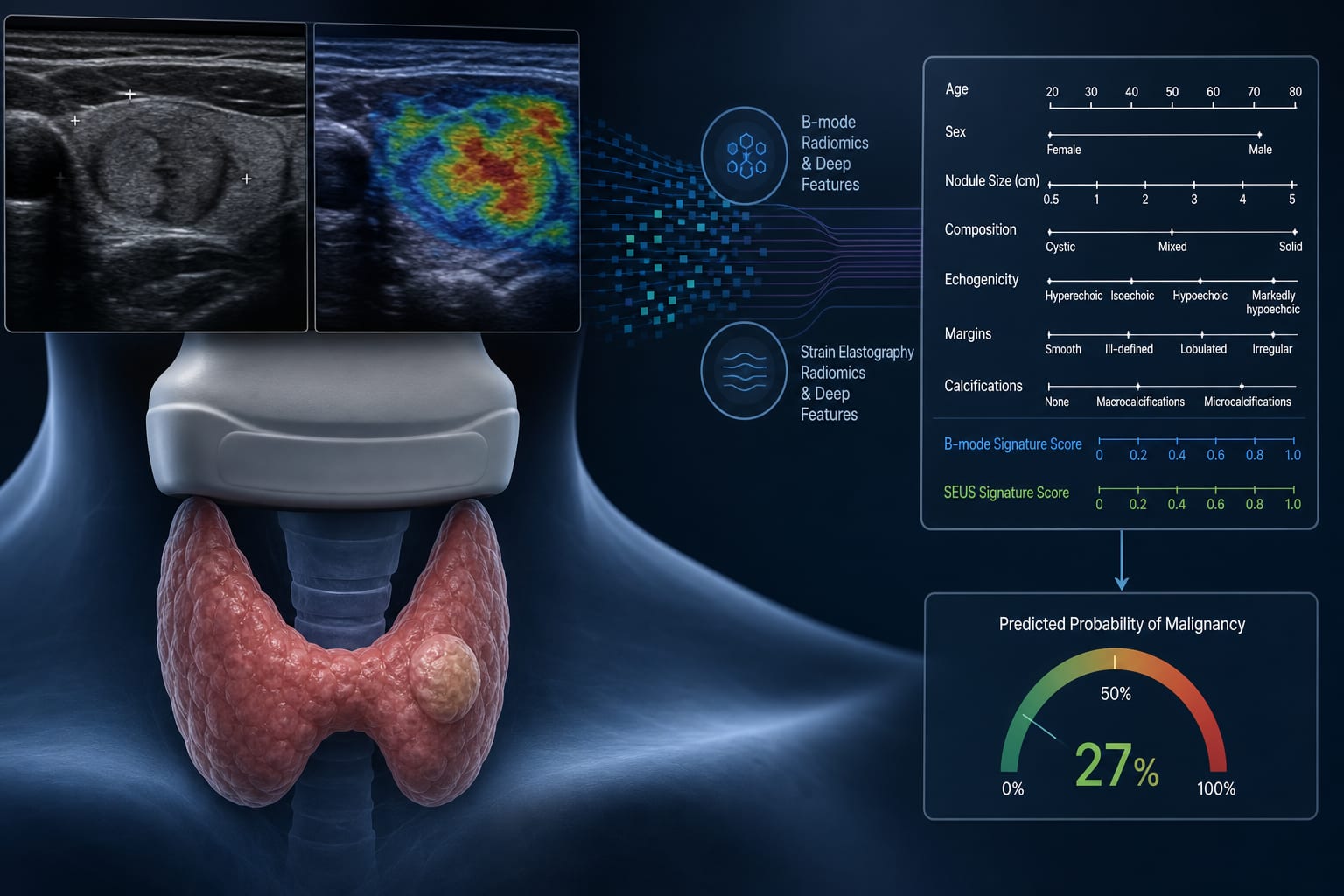
This study evaluates the performance of various deep learning models in distinguishing between benign and malignant thyroid nodules using multimodal ultrasound images.
Background
Thyroid cancer is a prevalent endocrine malignancy, making accurate differentiation between benign and malignant nodules crucial to avoid unnecessary procedures. Multimodal ultrasound techniques, such as superb microvascular imaging and shear-wave elastography, enhance imaging data but are subject to interpretation variability.
Data Highlights
Model
AUC
Accuracy
ResNet50
0.931
0.871
DenseNet121
0.857
-
VGG16
0.846
-
GoogLeNet
0.811
-
Key Findings
ResNet50 achieved the highest AUC of 0.931 in the validation cohort.
Deloong's test indicated ResNet50's AUC was significantly higher than other models (all P < 0.001).
ResNet50's accuracy (0.871) was better than junior radiologists (0.810) and comparable to intermediate radiologists (0.886).
Senior radiologists had the highest accuracy at 0.946.
Grad-CAM visualization showed ResNet50 focused on clinically relevant regions of thyroid nodules.
Clinical Implications
The findings indicate that deep learning models, particularly ResNet50, can enhance diagnostic accuracy for thyroid nodules.
Conclusion
The study demonstrates that multimodal ultrasound-based deep learning models can effectively differentiate between benign and malignant thyroid nodules.