Exo- and endoscopic two-step approach for recurrent vestibular schwannomas following surgical resection and radiosurgery: How I do it - Report - MDSpire
Combined Exo- and Endoscopic Technique for Recurrent Vestibular Schwannomas
Overview
This report details a two-step exo- and endoscopic approach (EETA) for managing recurrent vestibular schwannomas (VS) after prior surgery and radiosurgery. The technique enhances visualization and resection of intracanalicular and extracanalicular tumor components, improving outcomes in salvage surgery.
Background
Vestibular schwannomas often involve the internal auditory canal (IAC), where the fundus (FIAC) is divided into four quadrants housing critical nerves. The retrosigmoid approach (RSA) is commonly used for VS excision but has limited visualization of the FIAC. Recurrent tumors after surgery and radiosurgery pose challenges, necessitating techniques that allow clear observation of both the IAC and cerebellopontine angle (CPA) to optimize tumor removal and prevent recurrence.
Data Highlights
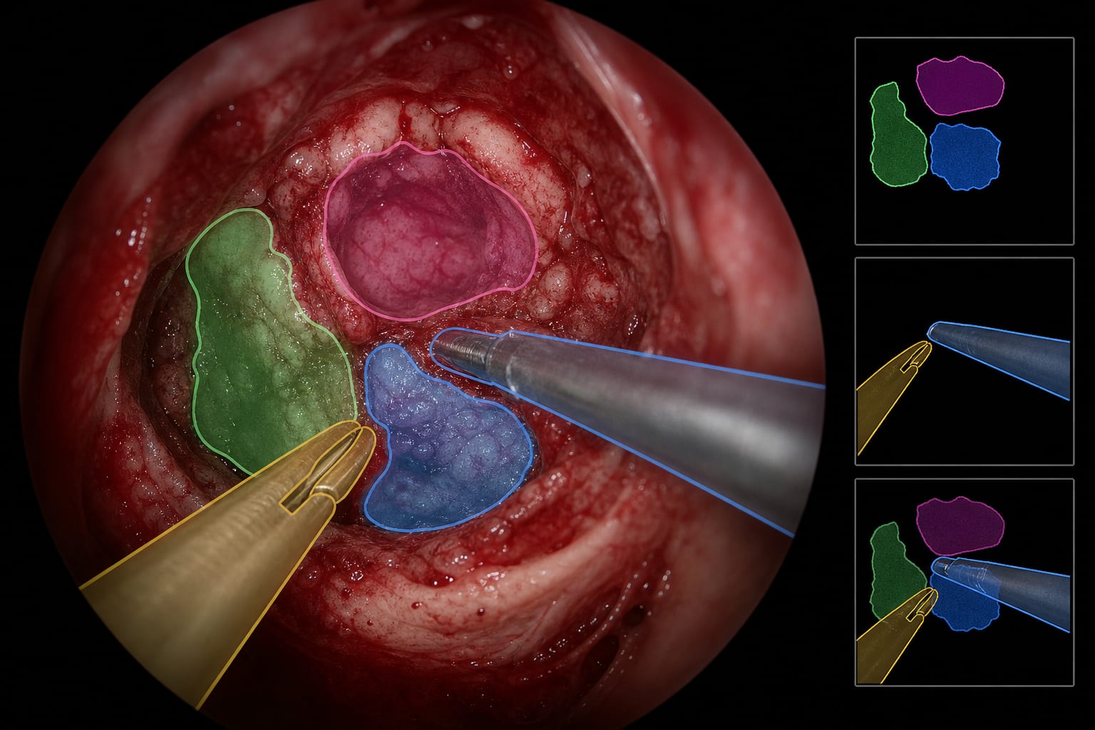
The described technique involves a two-step approach: first, removal of the extracanalicular tumor component using an exoscope, including limited intracanalicular tumor resection if visible; second, detailed inspection and resection of residual intracanalicular tumor using rigid endoscopes (0°, 30°, 70°), with the 70° endoscope enabling visualization of all four FIAC quadrants. Case 1 demonstrated successful salvage surgery 68 months after initial surgery with no facial paralysis and complete tumor removal confirmed by postoperative imaging.
Key Findings
The EETA technique combines exoscopic and endoscopic visualization to improve tumor resection in recurrent VS cases.
Use of a 70-degree endoscope allows comprehensive visualization of all FIAC quadrants, minimizing residual tumor risk.
Salvage surgery with EETA can be safely performed even after prior surgery and radiosurgery, with preservation of facial nerve function.
Sharp dissection under stereoscopic exoscopic visualization is critical for managing adhesions and scar tissue from previous treatments.
The approach facilitates removal of both extracanalicular and intracanalicular tumor components through a limited retrosigmoid craniotomy.
Clinical Implications
The combined exo- and endoscopic two-step approach offers a practical method to enhance visualization and resection of recurrent vestibular schwannomas, particularly in challenging salvage surgeries. Employing angled endoscopes, especially the 70-degree scope, aids in identifying residual tumor in the FIAC, potentially reducing recurrence rates while preserving facial nerve function.
Conclusion
The EETA technique represents a valuable advancement in the surgical management of recurrent vestibular schwannomas, enabling thorough tumor removal with minimal morbidity. Its application may improve long-term outcomes in patients requiring salvage surgery after prior interventions.
References
Kawase et al. -- Endoscopic Assistance in Vestibular Schwannoma Surgery
Pollock et al. -- Stereotactic Radiosurgery for Vestibular Schwannomas
Samii et al. -- Management of Recurrent Vestibular Schwannomas