Imaging Characteristics of Neonatal Horseshoe Lung Accompanied by Multiple Systemic Anomalies
Background
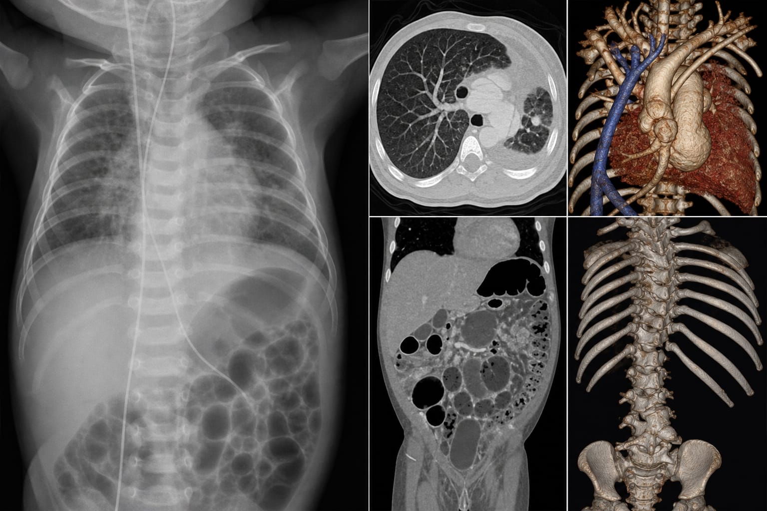
Horseshoe lung is a rare congenital pulmonary malformation. The coexistence of HL with other systemic anomalies, such as scimitar syndrome and VACTERL association, complicates clinical outcomes and necessitates thorough imaging for effective treatment planning.
Data Highlights
Imaging Modality
Findings
Cardiac Ultrasound
Patent ductus arteriosus, pulmonary hypertension, right pulmonary artery hypoplasia
Abdominal Ultrasound
Intestinal malrotation, abnormal intestinal loop distribution
Fusion of bilateral lower lungs, absence of typical bronchial branching
Key Findings
The patient presented with respiratory distress and multiple congenital anomalies.
Imaging revealed horseshoe lung with abnormal bronchial branching and pulmonary hypoplasia.
Associated conditions included scimitar syndrome and duodenal obstruction.
Multiple vertebral anomalies were identified, including butterfly vertebrae.
Clinical Implications
The case highlights the importance of using multiple imaging modalities to assess complex congenital anomalies in neonates. Early and accurate imaging can guide treatment decisions and improve prognostic assessments.
Conclusion
This case study enhances the understanding of horseshoe lung and its associated malformations, emphasizing the need for thorough imaging in managing such complex conditions.