Clinical Report: Vesicovaginal Reflux: A Case Study and Review
Overview
This report discusses a case of vesicovaginal reflux (VVR) in an 8-year-old girl, highlighting its underrecognition and the importance of dynamic imaging for diagnosis. A review of existing literature reveals a total of 58 cases, emphasizing the need for careful differentiation from anatomical abnormalities.
Background
Vesicovaginal reflux (VVR) is a functional voiding disorder that can lead to significant urinary incontinence, particularly in children. It is often misdiagnosed as anatomical issues like vesicovaginal fistula or obstructive hydrocolpos, which necessitate surgical intervention. Understanding VVR is crucial for appropriate management and to avoid unnecessary procedures.
Data Highlights
Measurement
Value
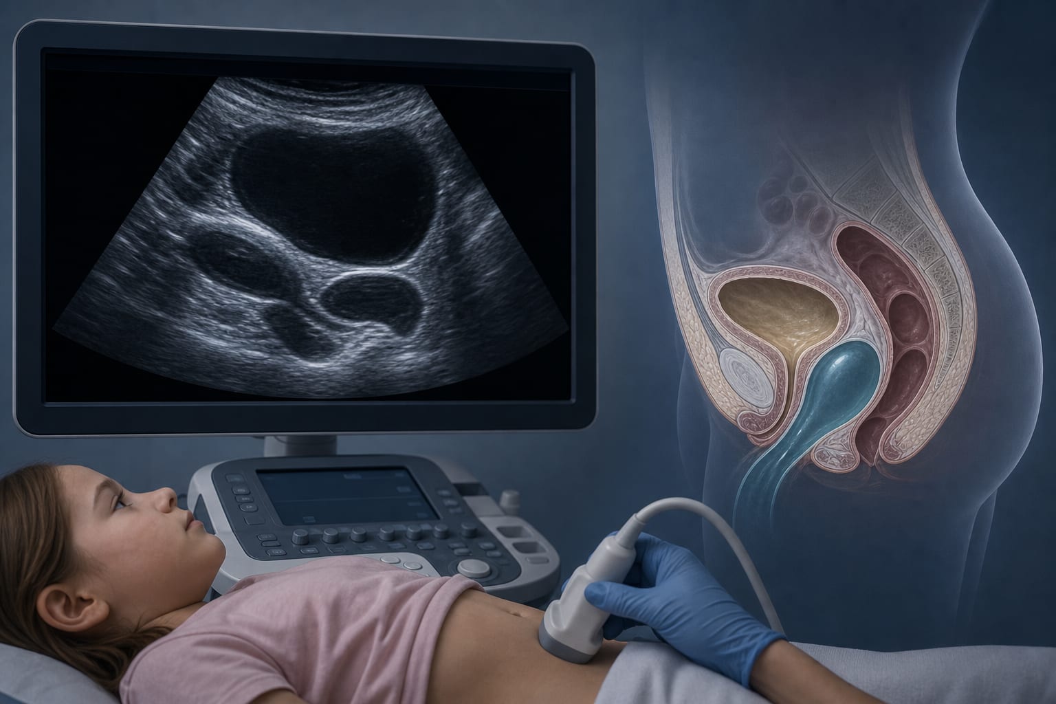
Anechoic area in vagina
7.3 × 3.3 × 4.0 cm
Body temperature
36.6°C
Height
142 cm
Weight
50 kg
BMI
24.79
Key Findings
VVR is characterized by retrograde urine flow into the vagina during micturition.
Dynamic imaging, particularly ultrasound, is essential for diagnosing VVR.
VVR can mimic anatomical abnormalities, leading to misdiagnosis.
Behavioral therapy is the primary treatment approach for VVR.
A total of 58 cases of VVR were reviewed, indicating its occurrence in various age groups.
Clinical Implications
Clinicians should consider VVR in cases of urinary incontinence, especially when imaging shows transient fluid collections in the vagina. Proper diagnosis can prevent unnecessary surgical interventions and guide effective management strategies focused on behavioral modifications.
Conclusion
Vesicovaginal reflux is an underrecognized condition that requires careful imaging and clinical evaluation for accurate diagnosis. Awareness and understanding of VVR can lead to better management and outcomes for affected patients.