Clinical Report: Examining the Clinicodermoscopic and Histopathological Progression of Acquired Reactive Perforating Collagenosis
Overview
This case study presents a 64-year-old woman with acquired reactive perforating collagenosis (ARPC) who experienced a misdiagnosis prior to the identification of early lesions. The findings highlight the importance of recognizing a pre-perforating stage in ARPC, which can aid in timely diagnosis and appropriate treatment.
Background
Acquired reactive perforating collagenosis is a rare condition often associated with systemic metabolic disorders such as diabetes mellitus. Its clinical presentation can lead to misdiagnosis, particularly before the characteristic transepidermal elimination becomes evident. Understanding the progression of ARPC is crucial for accurate diagnosis and management.
Data Highlights
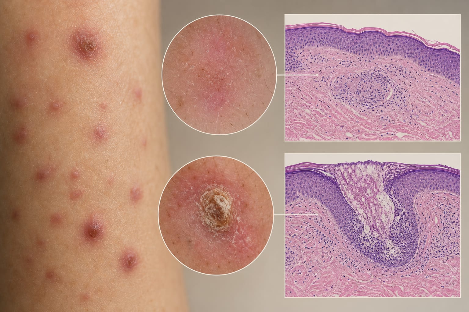
Case study of a 64-year-old woman with a 3-year history of pruritic papules and nodules, initially misdiagnosed as eczema. Dermoscopy revealed distinct features in early lesions, while biopsies showed abnormal collagen organization without transepidermal elimination.
Key Findings
The patient had a 3-year history of progressive pruritic papules and nodules.
Initial misdiagnosis as eczema led to ineffective treatments.
Dermoscopy of early lesions showed distinct features absent in fully developed lesions.
Biopsy of early lesions revealed abnormal collagen aggregation without transepidermal elimination.
Sequential histopathological analysis demonstrated a progression from superficial collagen remodeling to overt perforation.
Clinical Implications
Recognizing the pre-perforating stage of ARPC can facilitate timely diagnosis and appropriate biopsy selection, potentially reducing the risk of prolonged misdiagnosis. Clinicians should consider dermoscopy as a valuable tool in evaluating early lesions.
Conclusion
This case report provides direct evidence of a definable pre-perforating stage in ARPC, emphasizing the need for awareness of early signs to improve diagnostic accuracy.