Clinical Report: Comparing Intraoperative CBCT and Postoperative MDCT in Spine Surgery
Overview
This study compared image quality between intraoperative cone beam CT (CBCT) and postoperative multidetector CT (MDCT) in spine surgery patients. Findings suggest that CBCT provides comparable diagnostic image quality to MDCT, supporting its use for intraoperative assessment of spinal instrumentation.
Background
Intraoperative imaging in spine surgery has evolved from 2D fluoroscopy to 3D cone beam CT (CBCT), which enhances surgical precision and safety by allowing real-time assessment of implant placement. Postoperative multidetector CT (MDCT) remains the standard for confirming surgical outcomes. However, increased imaging raises concerns about cumulative radiation exposure, prompting evaluation of whether intraoperative CBCT can reduce the need for postoperative MDCT without compromising diagnostic quality.
Small (12.6×12.6 cm²), Medium (17.3×17.3 cm²), Large (25.2×19.5 cm²)
Customized per patient
Slice Thickness
0.45–0.49 mm
0.625 mm
Tube Voltage
120 kV fixed
100–120 kVp (thoracolumbar), 100 kVp (cervical)
Metal Artifact Reduction
Tilting C-arm ±20° to reduce artifacts
MAR algorithms when available
Key Findings
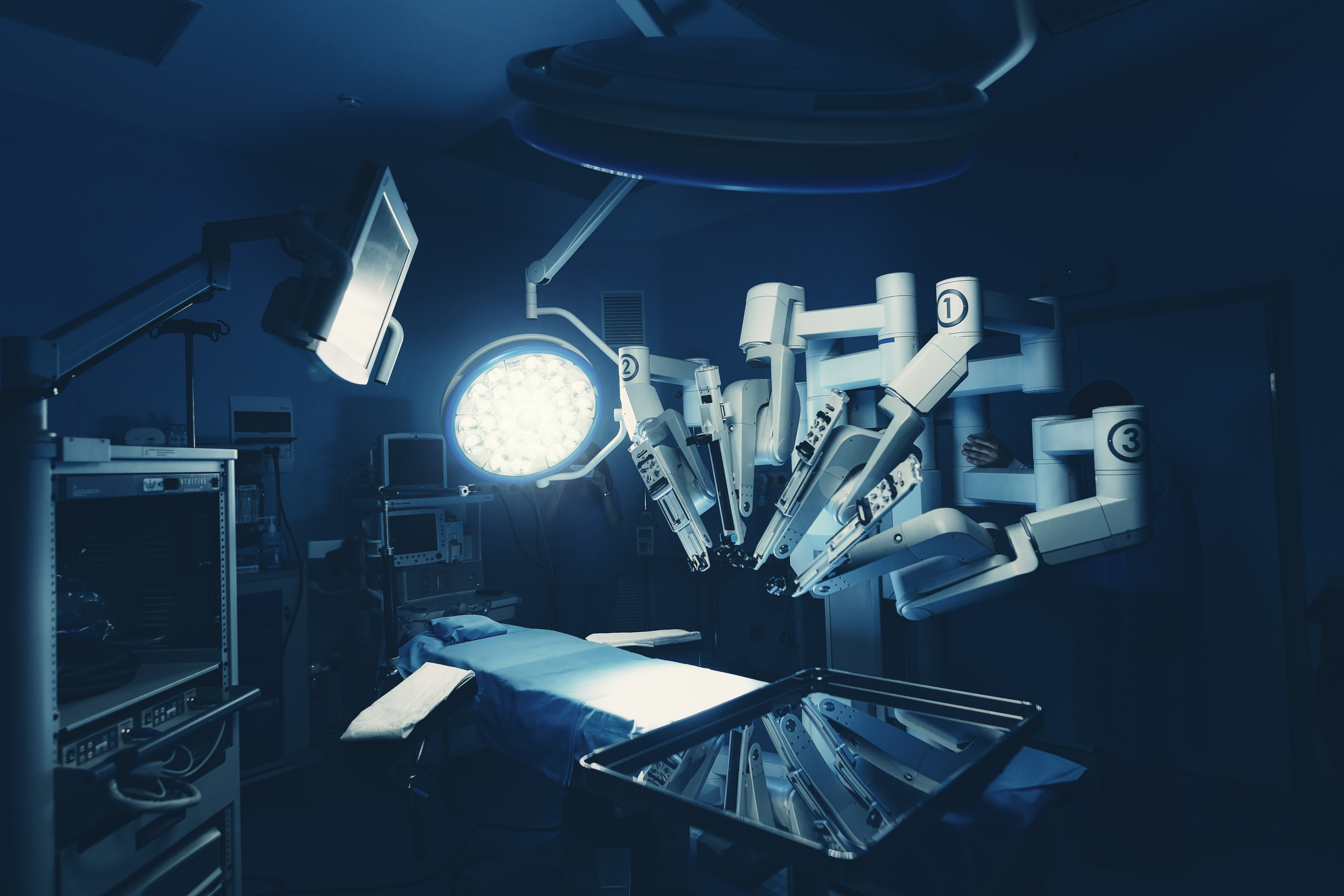
Intraoperative CBCT images were acquired immediately after implant placement using a ceiling-mounted robotic C-arm with 3D volumetric data capture.
Postoperative MDCT scans were performed with 128- or 256-row scanners using bone reconstruction algorithms and metal artifact reduction techniques.
207 paired CBCT and MDCT image sets were anonymized, randomized, and independently assessed for image quality.
CBCT demonstrated comparable image and diagnostic quality to MDCT for cervical and thoracolumbar spinal instrumentation.
CBCT protocols included variable field of view and slice thickness optimized for surgical level and patient size.
Use of CBCT intraoperatively has potential to reduce the need for postoperative MDCT, thereby lowering cumulative radiation exposure.
Clinical Implications
Intraoperative CBCT provides sufficient image quality to accurately assess spinal implants, potentially obviating the need for routine postoperative MDCT scans. This can streamline surgical workflows, reduce patient radiation dose, and optimize resource utilization. Clinicians should consider integrating CBCT into spine surgery protocols where available to enhance intraoperative decision-making and patient safety.
Conclusion
The study supports the use of intraoperative CBCT as a reliable imaging modality with diagnostic quality comparable to postoperative MDCT in spine surgery. Adoption of CBCT may reduce redundant imaging and associated radiation exposure without compromising clinical outcomes.
by Paulina Cewe, Mikael Skorpil, Alexander Fletcher-Sandersjöö, Victor Gabriel El-Hajj, Per Grane, Michael Fagerlund, Magnus Kaijser, Adrian Elmi-Terander, Erik Edström
Systematic review found robotic-assisted total hip arthroplasty improved implant positioning precision without demonstrating better patient-reported outcomes or lower complication rates than conventional surgery.