Intraoperative Evaluation of Sarcoma Surgical Margins With Indocyanine Green Fluorescence Imaging: Results From an Observational Study - Report - MDSpire
Clinical Report: Assessment of Surgical Margins in Sarcoma Procedures Using ICG
Overview
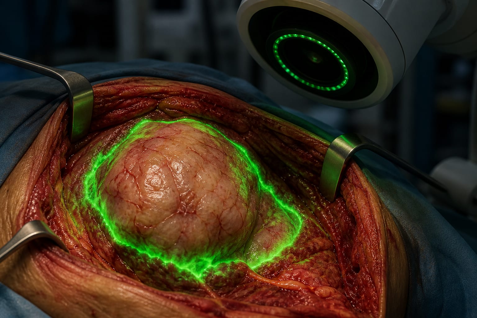
This observational study evaluates the use of indocyanine green (ICG) fluorescence imaging for assessing surgical margins in sarcoma surgeries. The findings indicate that a sarcoma-specific ICG protocol demonstrates high sensitivity in detecting positive margins.
Background
Sarcomas are rare but aggressive malignancies, with significant mortality rates despite advancements in treatment. Achieving negative surgical margins is critical in sarcoma management to minimize local recurrence, which can lead to severe complications. Current methods for margin assessment are often subjective.
Data Highlights
Parameter
Value
Number of patients
51
Previous study patients
26
ICG protocol sensitivity
High
Key Findings
ICG fluorescence imaging was evaluated in 51 patients, marking the largest report on its use in sarcoma surgery.
The new sarcoma-specific ICG protocol showed high sensitivity for detecting positive margins.
Previous imaging parameters from breast cancer surgery were adapted for sarcoma applications.
Local recurrence rates associated with positive surgical margins are reported in the literature.
Surgeons currently rely on subjective assessments for margin evaluation.
Clinical Implications
The use of ICG fluorescence imaging may enhance the accuracy of margin assessment during sarcoma surgeries.
Conclusion
The findings support the further investigation of ICG fluorescence imaging in sarcoma surgeries.
by Sumail Bhogal, Matthew Gong, William Li, Brenda Diergaarde, Alma Heyl, Clark Roth, Lawrence Garvin, Alexander Hoffman, Amanda Locker, Ines Lohse, Maria Silvaggio, Stella Lee, Richard McGough, Kurt Weiss