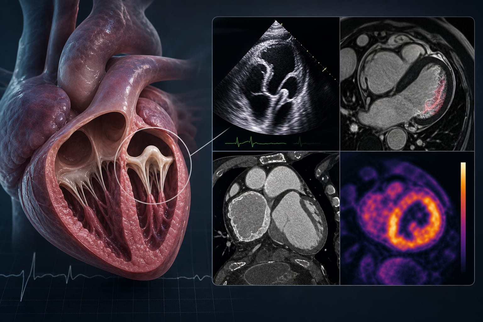
Multimodality cardiovascular imaging in arrhythmic mitral valve prolapse: a state-of-the-art review from structural assessment to myocardial tissue characterization
-
By
-
Giovanni Taverna
-
Kristian Galanti
-
Annagrazia Cecere
-
Francesca Graziano
-
Giancarlo Trimarchi
-
Roberta Antonazzo Panico
-
Antonella Cecchetto
-
Monica De Gaspari
-
Manuel Signorini
-
Aldo Carnevale
-
Giorgio De Conti
-
Raffaella Motta
-
Alberto Cipriani
-
Leonardo Calò
-
Fabrizio Ricci
-
Cristina Basso
-
C. Anwar A. Chahal
-
Martina Perazzolo Marra
-
June 15, 2026
-
0 min