Clinical Report: Retina Biomarkers for Treatment Selection in DME
Overview
Diabetic macular edema (DME) is a leading cause of vision loss, with a significant proportion of patients exhibiting resistance to anti-VEGF therapy. Emerging optical coherence tomography (OCT) biomarkers may enhance treatment stratification and guide individualized management strategies.
Background
DME affects millions globally and can lead to severe vision impairment if untreated. The shift from laser photocoagulation to anti-VEGF therapy has transformed DME management, yet approximately 40% of patients may not respond adequately to these treatments. Understanding the biological underpinnings of treatment resistance is crucial for optimizing patient outcomes.
Data Highlights
No numerical data available in the source material.
Key Findings
Approximately 19 million cases of DME are reported globally, projected to rise to 29 million by 2045.
Resistance to anti-VEGF therapy occurs in about 40% of DME patients, necessitating alternative treatment strategies.
OCT biomarkers, such as disorganization of retinal inner layers (DRIL), can predict visual acuity improvements with anti-VEGF therapy.
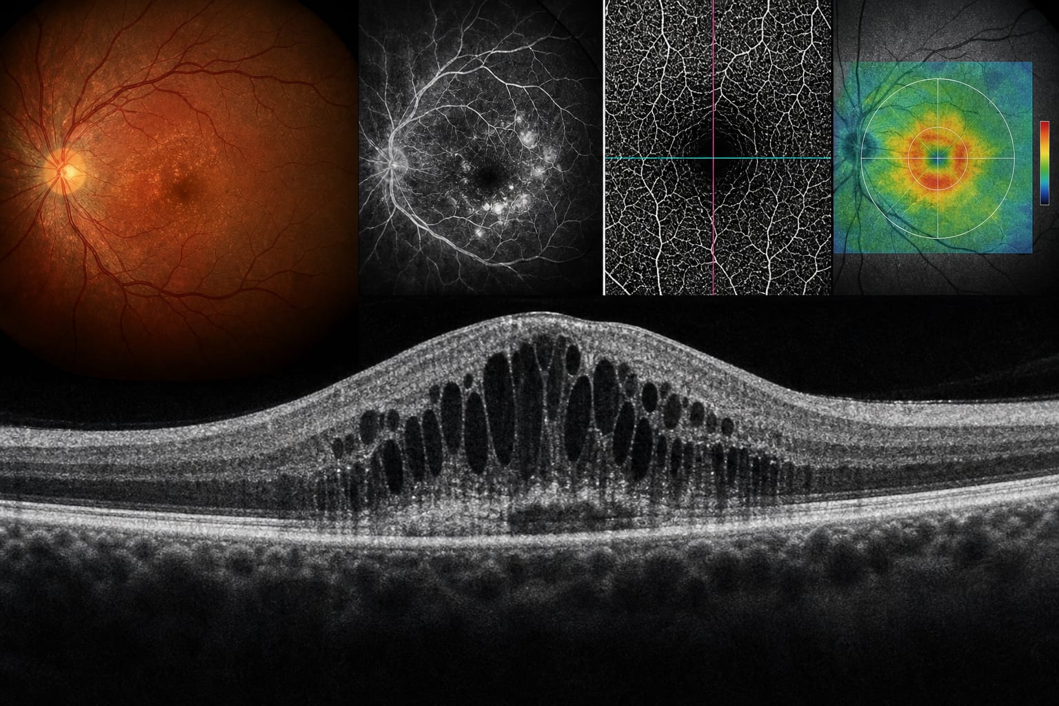
Presence of cystoid macular edema (CME) is associated with greater reductions in central subfield thickness (CST) after anti-VEGF treatment.
Multimodal imaging, including OCT and OCTA, is essential for comprehensive assessment and treatment planning in DME.
Clinical Implications
Clinicians should consider OCT biomarkers when assessing DME patients to tailor treatment strategies effectively. Early identification of patients likely to respond poorly to anti-VEGF therapy can facilitate timely alternative interventions, potentially improving visual outcomes.
Conclusion
The integration of OCT biomarkers into clinical practice may enhance the management of DME, allowing for more personalized treatment approaches and better patient outcomes.