Focal Atrial Tachycardia Arising from the Superior Vena Cava Post-Cryoablation
Overview
This case study describes a 58-year-old woman who developed focal atrial tachycardia originating from the superior vena cava following cryoablation for atrial fibrillation. High-resolution mapping and targeted radiofrequency applications successfully terminated the tachycardia.
Background
Focal atrial tachycardia can occur after catheter ablation for atrial fibrillation, often due to ectopic foci or incomplete ablation lines. Identifying the origin of these arrhythmias is crucial for effective treatment, particularly in patients with a history of prior ablation procedures. The superior vena cava is a recognized source of atrial ectopy that can lead to tachycardia.
Data Highlights
No numerical data or trial data available in the article.
Key Findings
A 58-year-old woman with recurrent paroxysmal atrial fibrillation developed focal atrial tachycardia post-cryoablation.
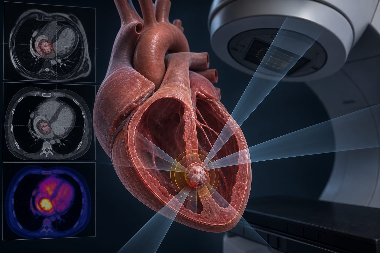
High-resolution 3D mapping identified the tachycardia's origin in the postero-septal part of the superior vena cava.
Three radiofrequency applications at the SVC level terminated the tachycardia and restored sinus rhythm.
During one year of follow-up, the patient remained asymptomatic with no arrhythmic recurrence.
Clinical Implications
High-resolution electrophysiological mapping is essential for identifying the origins of arrhythmias in patients with a history of atrial fibrillation ablation.
Conclusion
This case highlights the importance of precise mapping techniques in managing focal atrial tachycardia following prior ablation procedures. Successful intervention can lead to sustained sinus rhythm and improved patient outcomes.