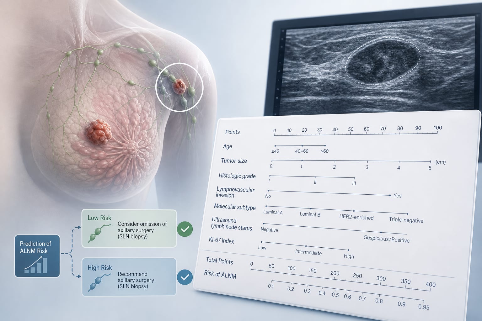
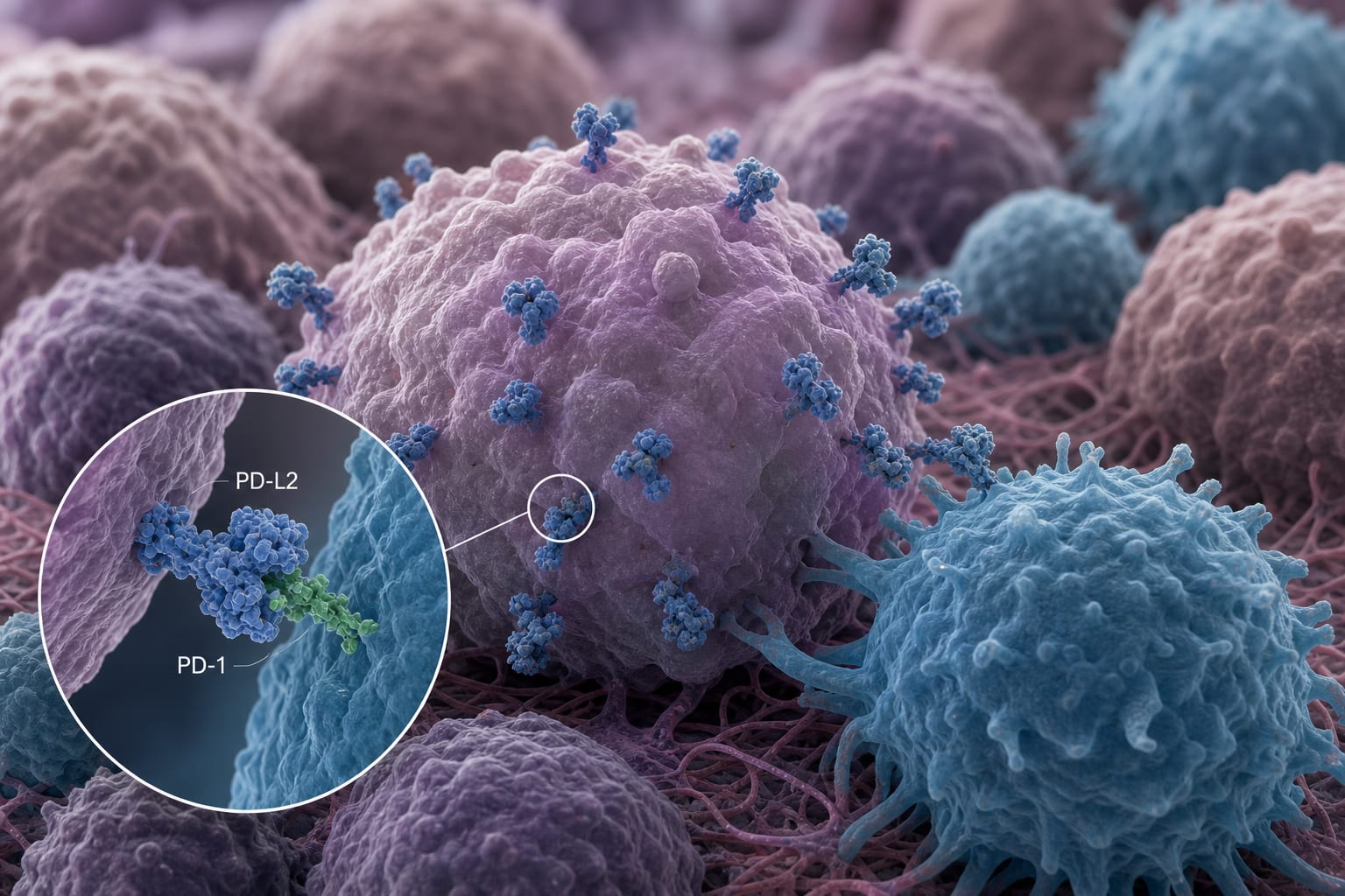
Technicolor Imaging Advances Cancer Immune Microenvironment Research
Overview
New spatial proteomics and transcriptomics technologies enable visualization of hundreds of markers simultaneously in tumor samples, revealing detailed immune cell locations and states. These advances help explain why some cancers, like triple-negative breast cancer, resist immunotherapy and chemotherapy.
Background
Traditional pathology techniques such as hematoxylin and eosin staining and immunohistochemistry have long been used to identify cells and proteins in tumor samples. However, these methods allow only limited multiplexing and lack spatial context for cell-to-cell interactions. Recent 'omics' approaches provide gene expression data but do not reveal the precise location of cells within the tumor microenvironment. Spatial proteomics and transcriptomics now allow researchers to visualize multiple proteins and RNA transcripts in situ, providing a comprehensive map of tumor and immune cell interactions.
Data Highlights
Spatial proteomics uses serial application of fluorescently labeled antibodies to detect every known protein on a single slide overnight. Spatial transcriptomics identifies RNA transcripts in each cell, revealing gene expression patterns with spatial resolution. These methods have been applied to triple-negative breast cancer models, differentiating active versus exhausted T cells and quiescent versus active cancer cells, highlighting immune exclusion zones within tumors.
Key Findings
- Traditional staining methods reveal limited markers and lack spatial resolution for cell interactions.
- Spatial proteomics enables simultaneous visualization of hundreds of proteins in tumor samples.
- Spatial transcriptomics complements proteomics by mapping RNA expression in situ.
- In triple-negative breast cancer, T cells cluster but cannot penetrate areas populated by dormant cancer cells, creating an immunological no-man's land.
- This spatial exclusion may explain resistance to immunotherapy and chemotherapy in certain tumors.
- These imaging advances provide new insights into tumor-immune dynamics and potential therapeutic targets.
Clinical Implications
Understanding the spatial distribution and functional states of immune and cancer cells within tumors can guide the development of more effective immunotherapies. Identifying immune-excluded tumor regions suggests new strategies to overcome resistance by targeting dormant cancer cells or modifying the tumor microenvironment. These technologies may eventually inform personalized treatment plans based on detailed tumor immune landscapes.
Conclusion
Technicolor spatial imaging technologies are transforming cancer research by revealing complex immune-tumor interactions previously hidden. This enhanced understanding holds promise for improving immunotherapy outcomes and developing novel therapeutic approaches.
References
- Wu C, Wucherpfennig K, Agudo J et al. (2023) Seeing Is Believing: How Technicolor Images of Tumor Samples are Changing Cancer Research
This content is an AI-generated, fully rewritten summary based on a published scholarly article. It does not reproduce the original text and is not a substitute for the original publication. Readers are encouraged to consult the source for full context, data, and methodology.