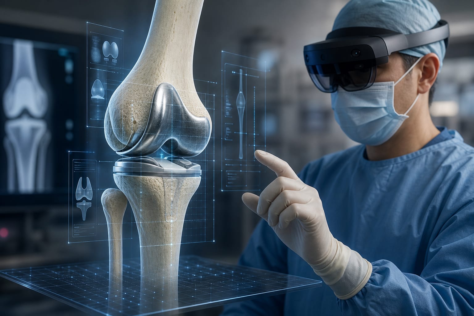
Clinical Report: Biomarkers and Imaging Indicators of Osteolysis Following TKA
Overview
This study identifies early risk factors for periprosthetic osteolysis after total knee arthroplasty (TKA) and establishes predictive biomarkers. Key findings indicate that leukocyte count, FIB-4 index, and uric acid-to-creatinine ratio are significant factors associated with osteolysis risk.
Background
Periprosthetic osteolysis is a major cause of failure after TKA, leading to complications such as prosthetic loosening and dislocation. With the increasing number of TKA procedures, understanding the risk factors for osteolysis is crucial for improving long-term outcomes and patient management. Identifying accessible biomarkers for early risk assessment can aid in the timely intervention and monitoring of patients post-surgery.
Data Highlights
Factor
Odds Ratio (OR)
95% Confidence Interval (CI)
P-value
Leukocyte Count
1.36
1.13–1.65
0.001
FIB-4 Index
1.94
1.20–3.14
0.007
Uric Acid-to-Creatinine Ratio
0.57
0.36–0.88
0.012
Key Findings
Leukocyte count is positively associated with osteolysis risk (OR = 1.36).
FIB-4 index shows a significant correlation with increased odds of osteolysis (OR = 1.94).
Higher uric acid-to-creatinine ratio is linked to lower odds of osteolysis (OR = 0.57).
Subgroup analyses indicate variations in risk factors based on sex, age, diabetes, and hypertension status.
Osteolysis is primarily driven by chronic inflammatory responses to wear debris from implants.
Clinical Implications
The identification of leukocyte count, FIB-4 index, and uric acid-to-creatinine ratio as risk factors for osteolysis can assist clinicians in early identification of high-risk patients. This may lead to improved management strategies and reduced postoperative complications.
Conclusion
The study highlights the importance of specific biomarkers in predicting periprosthetic osteolysis after TKA, which can facilitate early intervention and enhance patient outcomes.
With an aging population, spine disorders are becoming increasingly common. Age-related spinal degeneration is nearly universal, but not all patients experience symptoms—and not all degeneration progresses the same way.