Clinical Report: Enhancing glioblastoma studies through a brain slice model
Overview
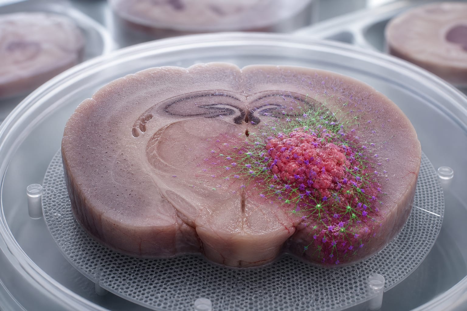
This study presents a novel whole murine brain slice model that maintains the complex cellular architecture of the brain, allowing for the investigation of glioblastoma (GBM) interactions with the tumor microenvironment. The model demonstrates viability for at least 7 days.
Background
Glioblastoma (GBM) is the most aggressive primary brain tumor in adults, characterized by rapid proliferation and diffuse infiltration. Traditional in vitro models often fail to replicate the complexity of human tissue and the tumor microenvironment (TME). There is a need for alternative models that reduce animal usage while maintaining physiological relevance for studying GBM biology and treatment responses.
Data Highlights
The study established a whole, large-format brain slice model from 6-9-day-old postnatal mice, allowing for the reduction of animal numbers by sectioning each brain into multiple slices. The slices were maintained in vitro for 7–14 days, serving as a carrier for GBM cell spheroids, and were assessed for viability using the AlamarBlue assay and live/dead staining methods.
Key Findings
The whole murine brain slice model preserves complex organotypic structures essential for GBM research.
The slices remained viable for a minimum of 7 days.
Co-culturing with GBM spheroids resulted in a slight decrease in slice viability due to tumor invasion.
Two fluorescence-based methods were employed to visualize and quantify tissue viability.
This model allows for examination of specific brain regions in relation to GBM.
Clinical Implications
The developed brain slice model provides a platform for studying GBM.
Conclusion
The establishment of this ex vivo whole-brain slice GBM model represents an advancement in glioblastoma research.