Case Report: Retained gallstone migrating to the chest wall after cholecystectomy presenting as chronic abscess, sinus tract, and inflammatory pseudotumor - Report - MDSpire
Advertisement
Case Study: Migration of a Retained Gallstone to the Chest Wall Following Cholecystectomy Resulting in Chronic Abscess, Sinus Tract, and Inflammatory Pseudotumor
Clinical Report: Migration of a Retained Gallstone to the Chest Wall
Overview
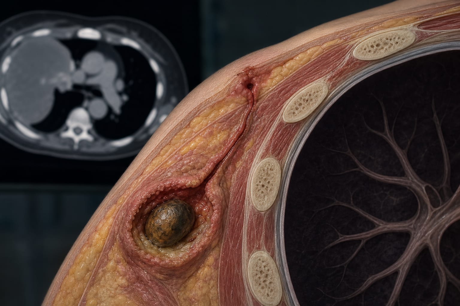
This case study presents a rare instance of a retained gallstone migrating to the chest wall two years post-cholecystectomy, resulting in a chronic abscess and inflammatory pseudotumor.
Background
Gallbladder perforation and stone spillage during laparoscopic cholecystectomy are common, occurring in up to 40% and 6%–10% of cases, respectively. While most retained stones remain asymptomatic, 2.3%–8.5% of patients may develop complications, including abscess formation and granuloma development. This case highlights the potential for retained gallstones to migrate to atypical locations, such as the chest wall.
Data Highlights
No numerical data or trial data presented in the article.
Key Findings
A 61-year-old woman developed a chronic abscess and sinus tract in the chest wall due to a retained gallstone.
CT imaging revealed a soft tissue mass with internal hyperdensity and rib erosion.
Histopathology confirmed chronic suppurative inflammation and features of an inflammatory pseudotumor.
The retained gallstone was found embedded in purulent material during surgical excision.
Complete surgical excision of the mass resulted in an uneventful recovery.
Clinical Implications
Imaging findings such as ring-like hyperdensity on CT and T2 hypointense nodules on MRI may indicate the presence of a retained gallstone.
Conclusion
This case illustrates the potential for retained gallstones to cause complications, including chronic chest wall pathology.