Clinical Report: Assessment of Haemodynamic Congestion via MRI and Its Impact on Clinical Outcomes
Overview
This study evaluates the use of MRI to assess haemodynamic congestion and its correlation with clinical outcomes in heart failure patients. MRI-derived measures, including pulmonary blood volume and left atrial volume, show promise in predicting adverse outcomes compared to traditional methods.
Background
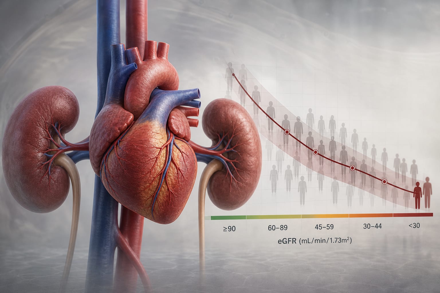
Heart failure is a significant global health issue, affecting over 25 million people and often linked to conditions like obesity and diabetes. Accurate assessment of haemodynamic congestion is crucial for effective diagnosis and management, as elevated left ventricular filling pressure is associated with poor prognosis. Traditional invasive methods like right heart catheterization are limited in routine clinical use, highlighting the need for non-invasive alternatives such as MRI.
Data Highlights
No numerical data available in the source material.
Key Findings
MRI-based physiological models integrating left atrial volume and left ventricular mass accurately estimate pulmonary artery wedge pressure.
These MRI measures outperform echocardiography in predicting clinical outcomes in heart failure patients.
Prolonged pulmonary transit time is associated with increased mortality and heart failure hospitalization.
Higher MRI-defined congestion correlates with myocardial fibrosis burden.
The study included 262 outpatients, demonstrating the feasibility of MRI in assessing congestion.
Clinical Implications
The findings suggest that MRI can serve as a valuable tool in the non-invasive assessment of haemodynamic congestion, potentially guiding treatment decisions in heart failure management. Clinicians may consider incorporating MRI-derived metrics into routine evaluations to enhance prognostic accuracy.
Conclusion
MRI offers a promising non-invasive alternative for assessing haemodynamic congestion in heart failure patients, with significant implications for clinical outcomes. Further validation in multicenter studies is warranted to establish its routine use in clinical practice.