Clinical Report: Fluoroscopic Metrics for Diagnosing Gastric Sleeve Stenosis
Overview
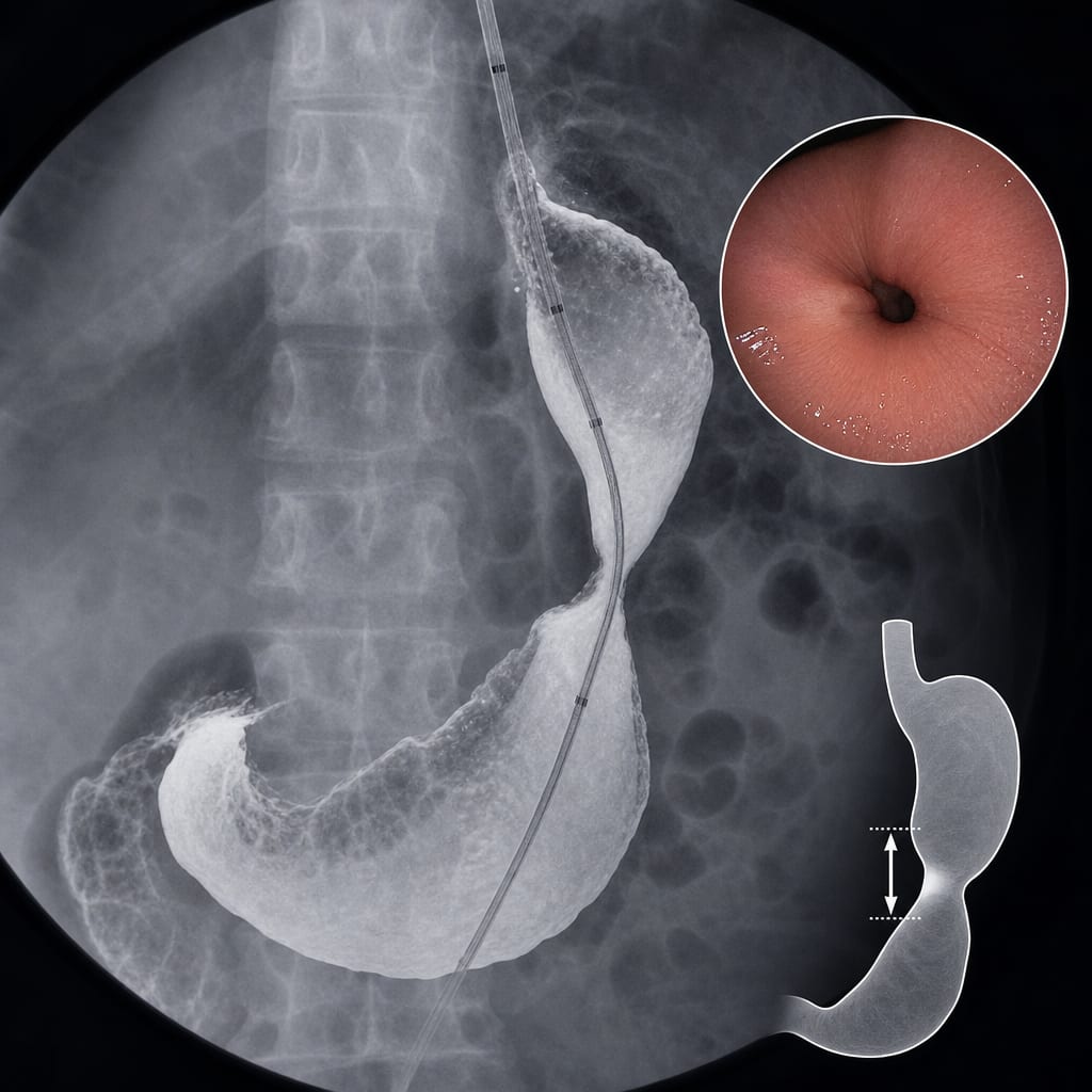
This report evaluates the use of fluoroscopy during upper endoscopy to diagnose and quantify gastric sleeve stenosis (GSS) in patients post-sleeve gastrectomy. The study highlights the potential for fluoroscopic metrics to provide objective parameters for assessing GSS severity, addressing a significant challenge in post-operative care.
Background
Sleeve gastrectomy is a widely performed bariatric procedure with a high efficacy for weight loss but carries risks of complications such as gastric sleeve stenosis. GSS can lead to obstructive symptoms and may affect patient outcomes if not diagnosed and managed appropriately. Current diagnostic methods lack sensitivity, necessitating the exploration of more reliable techniques like fluoroscopy.
Data Highlights
No numerical data available in the source material.
Key Findings
Gastric sleeve stenosis occurs in 1–4% of patients post-sleeve gastrectomy.
Symptoms of GSS include regurgitation, dyspepsia, and abdominal pain.
Fluoroscopy can provide objective metrics for diagnosing and quantifying GSS severity.
Endoscopy remains crucial for assessing luminal narrowing and planning treatment.
Automated image analysis and impedance planimetry may enhance diagnostic accuracy but are not widely available.
Clinical Implications
The integration of fluoroscopy into endoscopic evaluations may improve the diagnosis and management of gastric sleeve stenosis. Clinicians should consider this technique to enhance the objectivity of GSS assessments and guide treatment decisions effectively.
Conclusion
Fluoroscopy represents a promising tool for diagnosing gastric sleeve stenosis, potentially improving patient outcomes through more accurate assessments. Further research is needed to validate its effectiveness in routine clinical practice.