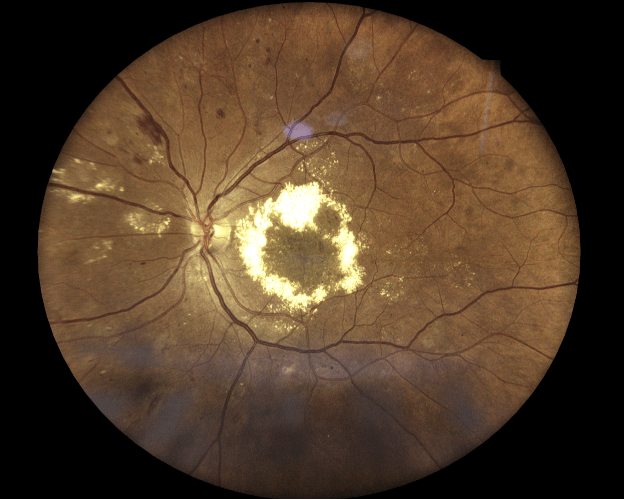
Clinical Report: Neo or Not Neo? Widefield OCT-A Reveals Hidden Neovascularization
Overview
Widefield OCT and OCT-A effectively identify hidden retinal neovascularization in patients with complex retinopathy, emphasizing the importance of advanced imaging in diagnosing proliferative diabetic and sickle cell retinopathy.
Background
The detection of retinal neovascularization is crucial in managing multifactorial retinopathy, particularly in diabetic patients. Early identification can significantly impact treatment decisions and patient outcomes. Advanced imaging techniques like OCT and OCT-A enhance diagnostic capabilities, allowing for better assessment of retinal conditions.
Data Highlights
No numerical data provided in the article.
Key Findings
Widefield OCT and OCT-A can uncover hidden neovascularization in complex retinopathy cases.
The patient presented with bilateral blurred vision and a significant medical history including diabetes and sickle cell trait.
OCT-A confirmed the diagnosis of proliferative diabetic and sickle cell retinopathy.
Advanced imaging technologies improve diagnostic accuracy and clinical decision-making.
Utilization of OCT-A is essential for assessing macular edema associated with retinal neovascularization.
Clinical Implications
Clinicians should consider incorporating widefield OCT and OCT-A in routine evaluations of patients with diabetes and other risk factors for retinal disease, following specific protocols to enhance detection of neovascularization and inform treatment strategies.
Conclusion
The use of advanced imaging techniques like OCT-A is vital for accurate diagnosis and management of retinal diseases, particularly in patients with multifactorial retinopathy, as early detection can significantly improve patient outcomes.