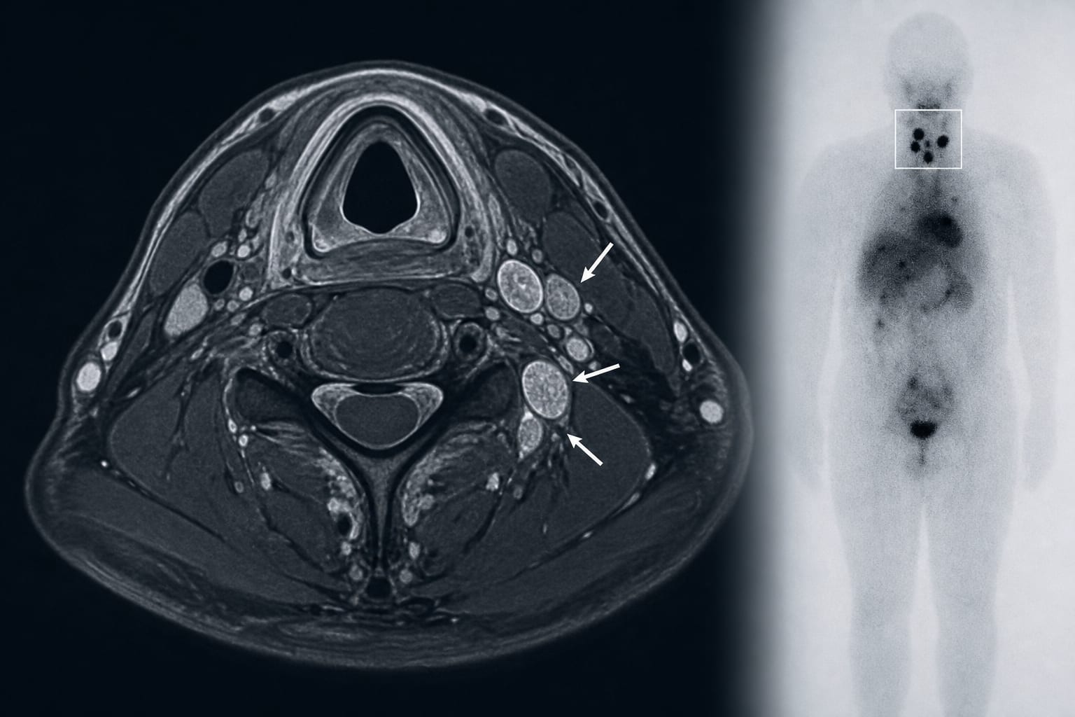
MRI characteristics of cervical radioiodine-avid lymph nodes detected on post-therapeutic ¹³¹I whole-body scintigraphy in differentiated thyroid carcinoma
-
By
-
Liang-Qian Tong
-
Zhuo-Wen Li
-
Wei Liu
-
Yan-Fang Sui
-
Yue Chen
-
June 23, 2026
-
0 min