Retinal Imaging Model Enhances Detection of Coronary Artery Disease
Overview
A retinal imaging–based model demonstrated moderate-to-good accuracy in detecting coronary artery disease (CAD), outperforming clinical risk factors alone. Combining retinal features with clinical variables improved diagnostic performance in a retrospective study of patients undergoing coronary angiography.
Background
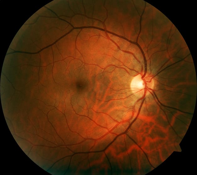
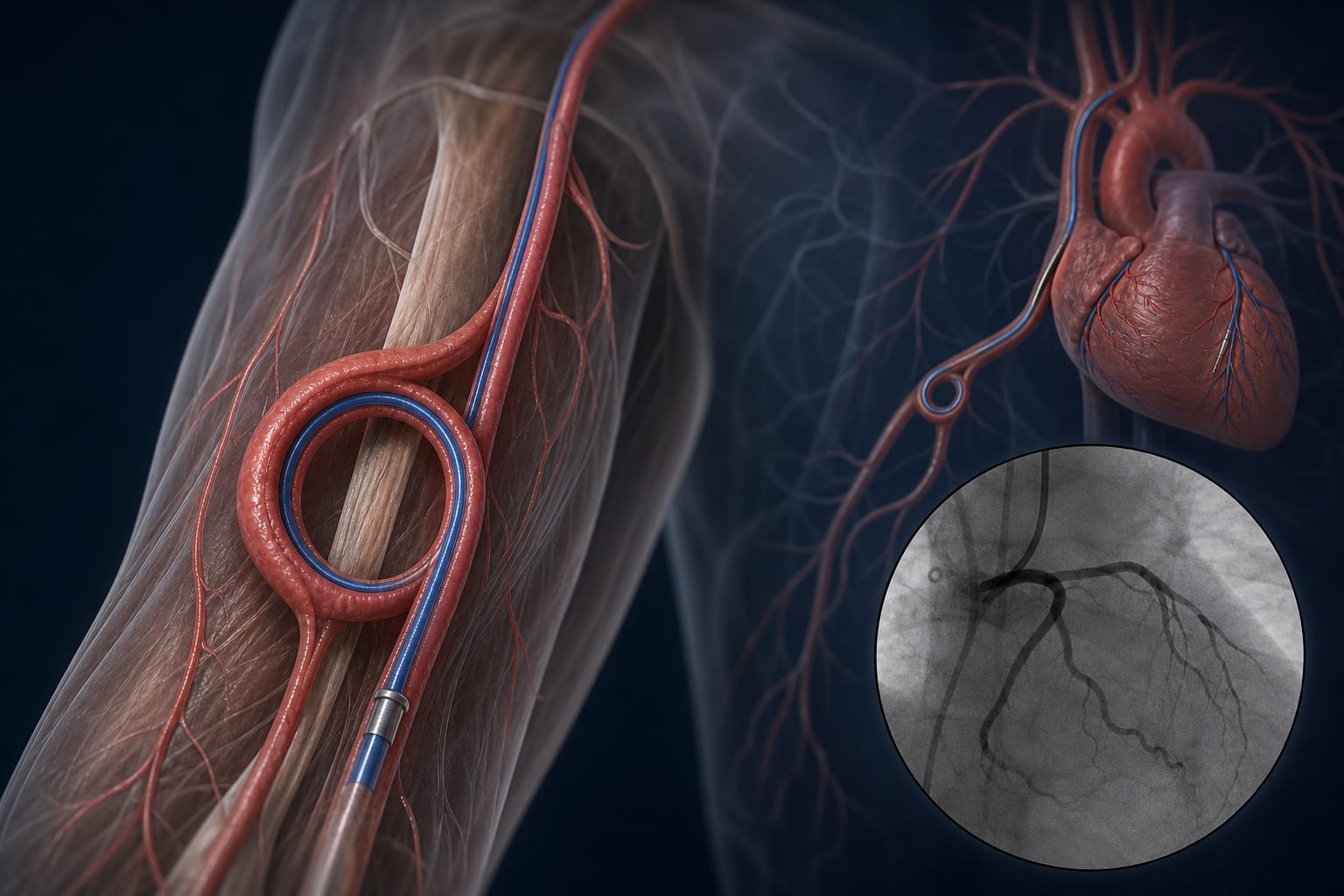
Coronary artery disease remains a leading cause of morbidity and mortality worldwide, necessitating improved risk assessment tools. The retinal microvasculature shares structural and pathophysiological characteristics with coronary vessels, offering a noninvasive window into systemic vascular health. Retinal imaging has emerged as a promising modality for cardiovascular risk evaluation, with quantified retinal vascular parameters potentially serving as biomarkers for CAD. This study evaluated the diagnostic utility of retinal imaging combined with clinical risk factors in a high-risk Chinese cohort.
Data Highlights
Model
AUROC
Sensitivity
Specificity
Combined retinal imaging + clinical factors
0.802
0.797
0.679
Clinical risk factors alone
0.748
Not reported
Not reported
Retinal imaging alone
0.694
Not reported
Not reported
Key Findings
Quantitative retinal vascular parameters such as fractal dimension and vessel density were independently associated with CAD after adjustment for confounders.
In the combined model, decreased fractal dimension, reduced optic disc axis ratio, and shorter optic disc-to-macula distance were retained as independent retinal predictors.
Clinical predictors included male sex, dyslipidemia, and elevated lipoprotein(a), triglycerides, LDL cholesterol, and glycated hemoglobin levels.
The combined model achieved an AUROC of 0.802, with sensitivity of 79.7% and specificity of 67.9%, outperforming models using clinical or retinal data alone.
The model demonstrated higher sensitivity than specificity, indicating greater utility for identifying patients at increased CAD risk rather than ruling out disease.
The study used non-mydriatic fundus photography and explicitly quantified retinal features, enhancing interpretability and potential clinical feasibility.
Clinical Implications
Retinal vascular phenotyping may serve as a valuable adjunct to traditional clinical risk assessment for CAD, particularly when combined with established risk factors. The use of noninvasive retinal imaging could facilitate earlier identification of high-risk patients in clinical practice. However, clinicians should consider the current evidence is limited to a high-risk, predominantly Han Chinese population and requires further validation before widespread adoption.
Conclusion
This study supports the potential of retinal imaging combined with clinical variables to improve CAD detection. Further research in larger, diverse cohorts is necessary to confirm these findings and establish broader clinical utility.
Related Resources & Content
BMJ Open -- Retinal Imaging Model Aids CAD Detection
In a pooled analysis of two randomized crossover trials, reducing nightly sleep by about 1.5 hours for 6 weeks was associated with modest increases in body weight and waist circumference without measurable changes in body composition.