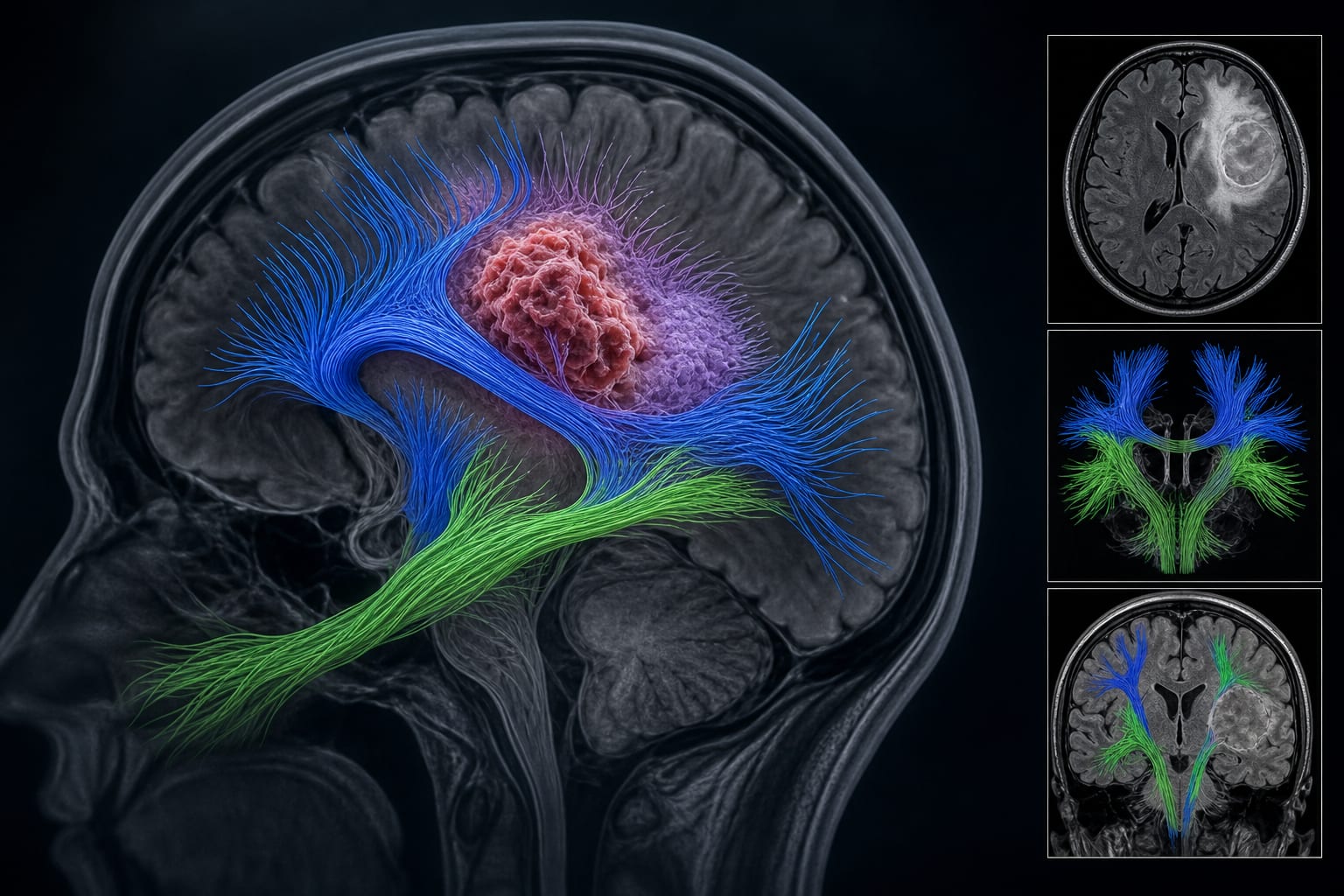
Combining Deep Learning and Thermal Analysis to Enhance MRI Brain Tumor Diagnosis
Overview
This study introduces a MATLAB-based AI pipeline integrating deep learning segmentation, classification, and thermal estimation to improve MRI brain tumor diagnosis. By combining grayscale MRI data with thermal pseudo-coloring and texture analysis, the model enhances malignancy prediction and physiological tumor characterization.
Background
Brain tumors present significant diagnostic challenges due to their heterogeneity in type, size, and location. MRI remains the gold standard for brain tumor detection but relies heavily on radiologist expertise and grayscale imaging. Recent advances in AI, particularly convolutional neural networks and vision transformers, have improved tumor classification and segmentation accuracy. However, these methods often overlook physiological tumor characteristics such as temperature variations, which can indicate tumor aggressiveness.
Data Highlights
Metric
Value
Classification Accuracy
High (exact value not specified)
Precision
Reported (exact value not specified)
Recall
Reported (exact value not specified)
F1-Score
Reported (exact value not specified)
Intersection over Union (IoU)
Reported (exact value not specified)
Key Findings
A unified MATLAB AI pipeline was developed combining tumor segmentation (U-Net), classification (CNN), thermal estimation, and malignancy grading.
Tumor temperature was estimated using a logarithmic function of tumor area (mm²) and visualized with thermal pseudo-coloring.
GLCM texture features (contrast, correlation, energy, homogeneity) were effective in malignancy classification and correlated with tumor size and temperature.
Thermal overlays enhanced tumor annotation and physiological modeling beyond traditional grayscale MRI analysis.
The integrated approach addresses limitations of prior models that relied solely on grayscale MRI without physiological context.
Clinical Implications
Incorporating thermal analysis with AI-driven MRI interpretation may improve early detection and malignancy assessment of brain tumors by providing additional physiological insights. This approach could reduce reliance on subjective radiologist interpretation and enhance diagnostic accuracy and efficiency. The methodology supports non-invasive tumor characterization, potentially guiding treatment planning and prognosis.
Conclusion
This study demonstrates that combining deep learning with thermal imaging and texture analysis enhances MRI-based brain tumor diagnosis by integrating anatomical and physiological data. The proposed pipeline offers a promising tool for improved tumor detection and malignancy grading in neuro-oncology.
Related Resources & Content
Aleid et al. -- Deep Learning for MRI Brain Tumor Classification
Li -- AI in Tumor Segmentation and Classification
Kaifi -- Role of AI in Clinical Decision-Making
Bousselham et al. -- Brain Tumor Impact on Local Temperature Distribution
Owens et al. -- Metabolic Heat Production Simulation in Tumors
Ring and Ammer -- Infrared Imaging for Non-Invasive Diagnosis
Over two days, specialists across neurology, neurosurgery and related subspecialties came together to discuss advances in stroke care, epilepsy, movement disorders, neurodegenerative disease, neuro-oncology, brain and spine surgery, interventional pain management and emerging technologies.