Clinical Report: Streamlined DeepME Framework for Macular Edema Management
Overview
The DeepME framework, utilizing an enhanced YOLOv11 architecture, demonstrates high accuracy in detecting macular edema in OCT images.
Background
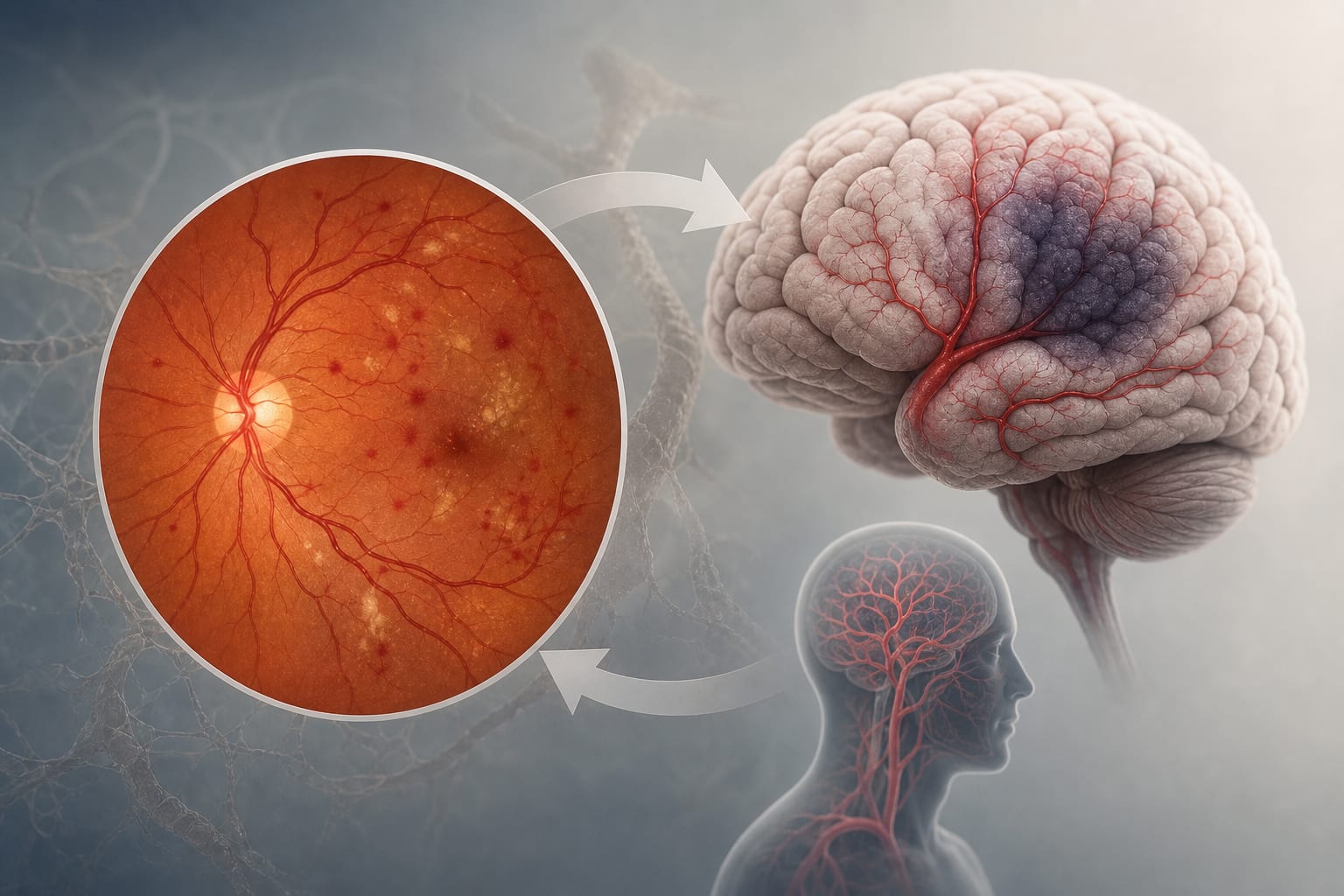
Macular edema is a significant complication of diabetic retinopathy and retinal vein occlusion, leading to vision impairment in millions globally. Current treatments, primarily anti-VEGF injections, often face challenges with patient response variability and persistent edema. The integration of advanced imaging techniques like OCT with artificial intelligence is being explored.
Data Highlights
Metric
DeepME Performance
Accuracy
0.980
Specificity
0.990
Sensitivity
0.970
Precision
0.990
F1-score
0.980
AUC
0.9993
Key Findings
DeepME achieved an accuracy of 0.980 and an AUC of 0.9993 in macular edema detection.
Significant reduction in central foveal thickness was observed in the anti-VEGF cohort at one-month follow-up (p < 0.001).
DeepME demonstrated substantial agreement with manual grading (p < 0.001).
The framework integrates clinical guidelines and expert knowledge for treatment recommendations.
Grad-CAM visualizations confirmed precise localization of cystoid macular edema.
Clinical Implications
DeepME provides a robust tool for clinicians to enhance the accuracy of macular edema diagnosis and treatment planning. Its integration of clinical guidelines may streamline decision-making processes in managing patients with diabetic retinopathy and retinal vein occlusion.
Conclusion
The introduction of DeepME represents a significant advancement in the management of macular edema, offering high accuracy and comprehensive support for clinical decision-making.