Late recurrence and histological progression of a cervical intradural extramedullary solitary fibrous tumor after gross total resection: a case report - Report - MDSpire
Advertisement
Late recurrence and histological progression of a cervical intradural extramedullary solitary fibrous tumor after gross total resection: a case report
Clinical Report: Delayed Recurrence of Solitary Fibrous Tumor in Cervical Space
Overview
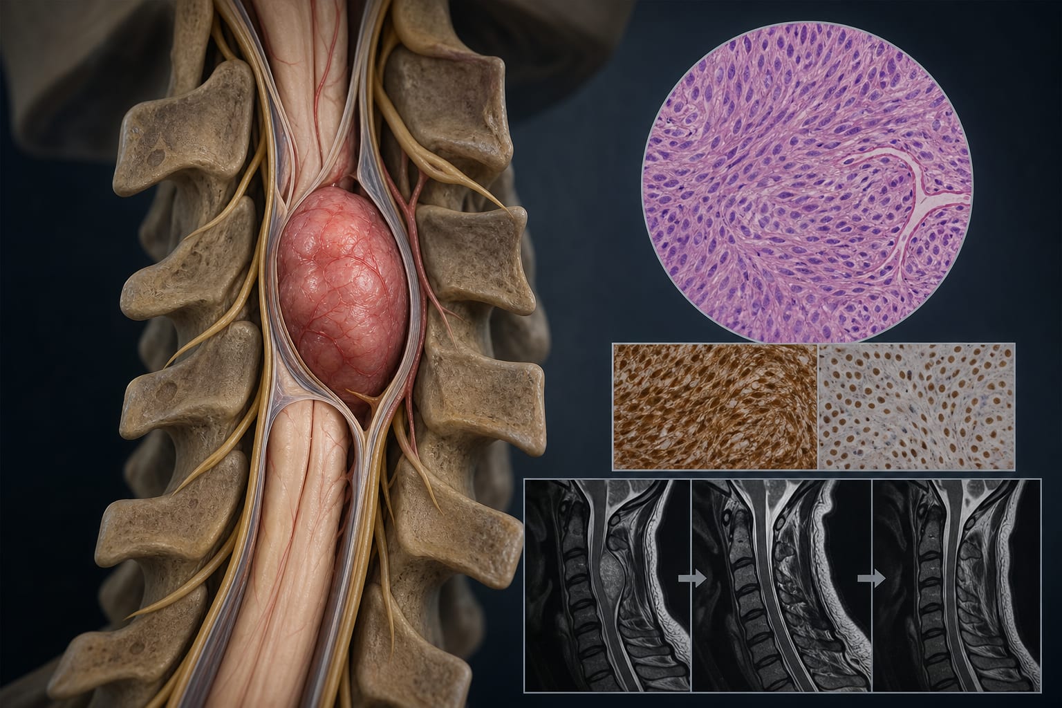
This case study presents a 39-year-old male with a solitary fibrous tumor (SFT) in the cervical intradural extramedullary space, highlighting the potential for delayed recurrence and histological progression even after gross total resection. The patient experienced a recurrence five years post-surgery, with histological advancement from WHO grade 2 to grade 3.
Background
Solitary fibrous tumors are rare mesenchymal neoplasms of the central nervous system, often exhibiting unpredictable biological behavior. Spinal localization is uncommon, particularly in the cervical intradural extramedullary space, making long-term outcomes and management strategies poorly defined. Understanding the potential for delayed recurrence is crucial for optimizing patient surveillance and treatment.
Data Highlights
No numerical data or trial data presented in the article.
Key Findings
The patient underwent gross total resection of a cervical SFT with initial favorable outcomes.
Five years post-surgery, the patient developed recurrent symptoms and MRI revealed a localized lesion at the surgical site.
Histopathological examination of the recurrence showed increased mitotic activity and necrosis, indicating progression to WHO grade 3.
Close radiological surveillance was recommended following the second surgery.
Delayed recurrence and histological progression can occur even after complete resection of initially low-grade tumors.
Clinical Implications
This case underscores the importance of long-term radiological follow-up for patients with solitary fibrous tumors, even after gross total resection. Clinicians should consider multidisciplinary evaluations for recurrent or higher-grade tumors to determine appropriate adjuvant treatment strategies.
Conclusion
The findings from this case highlight the need for ongoing surveillance in patients with solitary fibrous tumors due to the risk of delayed recurrence and histological progression, emphasizing the complexity of managing these rare neoplasms.
by Yergen N. Kenzhegulov, Daniyar K. Zhamoldin, Viktor G. Aleinikov, Talgat T. Kerimbayev, Berik Zhetpisbaev, Makar P. Solodovnikov, Aisa Z. Nurpeisov, Serik Akshulakov