Clinical Report: Utilizing Indocyanine Green Fluorescence Imaging for Resection of Complex FNH
Overview
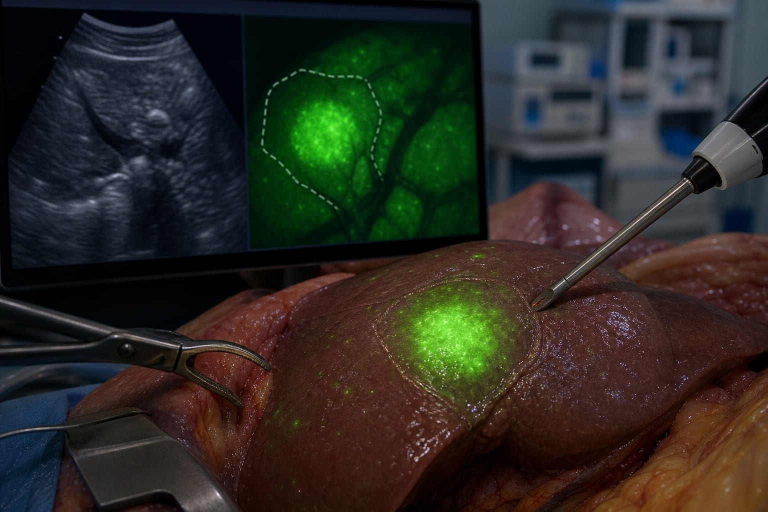
This report presents two cases where indocyanine green (ICG) fluorescence imaging was used in conjunction with intraoperative ultrasonography for laparoscopic excision of focal nodular hyperplasia (FNH). Both patients achieved successful wedge excision with negative surgical margins, highlighting the potential of ICG in enhancing surgical precision.
Background
Focal nodular hyperplasia (FNH) is a common benign liver lesion often managed conservatively. However, atypical presentations may necessitate surgical intervention for definitive diagnosis or symptom management. The integration of ICG fluorescence imaging with intraoperative ultrasonography may improve surgical outcomes in complex cases involving perivascular lesions.
Data Highlights
Patient
Lesion Size (cm)
Estimated Blood Loss (mL)
Surgical Outcome
Case 1
3.3 x 2.1
50
Negative margins
Case 2
Not specified
20
Negative margins
Key Findings
ICG fluorescence imaging combined with IOUS provided clear visual contrast for lesion delineation.
Both patients underwent successful wedge excision with negative surgical margins.
Estimated blood loss was minimal, ranging from 20 to 50 mL.
Incidental hyperfluorescent foci were noted, with one showing benign hyperplastic changes.
The clinical significance of remaining hyperfluorescent foci is uncertain.
Clinical Implications
The use of ICG fluorescence imaging may enhance the precision of laparoscopic resections for complex FNH lesions, particularly those located near major vascular structures. Further studies are warranted to evaluate the reproducibility and clinical value of this technique.
Conclusion
The integration of ICG fluorescence imaging with intraoperative ultrasonography appears to facilitate effective surgical management of complex FNH cases, though additional research is needed to confirm its broader applicability.
For many people, cardiovascular disease develops silently. It can remain undetected for years, overshadowed by an active lifestyle and an apparent lack of symptoms.