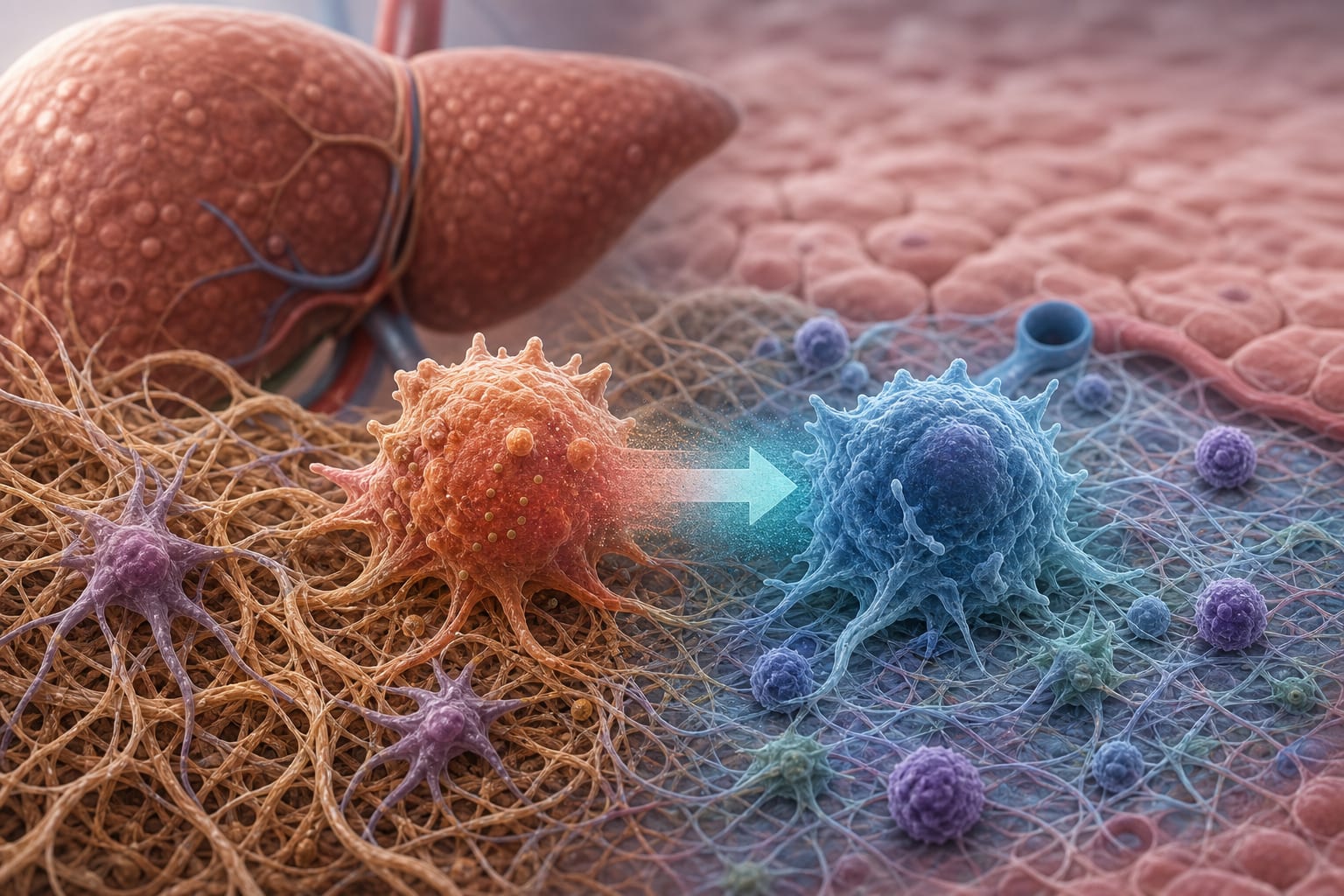
Integrating multi-omics and machine learning systematically deciphers cellular heterogeneity and fibrotic regulatory networks in the progression from MASLD to MASH - Report - MDSpire
Advertisement
Integrating multi-omics and machine learning systematically deciphers cellular heterogeneity and fibrotic regulatory networks in the progression from MASLD to MASH
Multi-Omics and Machine Learning Reveal Cellular Drivers of MASLD to MASH Progression
Overview
This study integrates multi-omics datasets and machine learning to identify key cellular changes and regulatory pathways driving the progression from MASLD to MASH. A distinct DTNA+ macrophage subpopulation and the RUNX2–PLG–PARD3 signaling axis were implicated in liver fibrosis and serve as potential diagnostic and therapeutic targets.
Background
Metabolic dysfunction-associated steatotic liver disease (MASLD) can progress to metabolic dysfunction-associated steatohepatitis (MASH), a critical step leading to cirrhosis and hepatocellular carcinoma. The cellular mechanisms underlying this transition remain poorly understood. Recent advances in single-cell and spatial transcriptomics enable detailed mapping of liver microenvironment remodeling. Machine learning approaches can further identify biomarkers distinguishing disease stages and reveal novel therapeutic targets.
Data Highlights
Parameter
Finding
Major liver cell types identified
7
Cell types enriched in MASH
Monocytes/macrophages and hepatic stellate cells
Distinct macrophage subpopulation
DTNA+ macrophages enriched in MASH
Machine learning model performance (mean AUC)
0.839
Key transcriptional regulator
RUNX2
Key Findings
Seven major liver cell types were identified, with monocytes/macrophages and hepatic stellate cells significantly enriched and spatially co-localized in MASH.
A distinct DTNA+ macrophage subpopulation was specifically enriched in MASH, exhibiting M2 polarization, hypoxia, and enhanced inflammatory signaling.
Pseudotime trajectory analysis indicated that DTNA+ macrophages differentiate from Kupffer cells, with RUNX2 as a key transcriptional regulator.
Cell communication analysis revealed interaction between DTNA+ macrophages and activated hepatic stellate cells via the RUNX2–PLG–PARD3 axis, promoting liver fibrosis.
Ensemble machine learning models identified DTNA as the optimal biomarker to distinguish MASLD from MASH with a mean AUC of 0.839.
Clinical Implications
The identification of DTNA+ macrophages and the RUNX2–PLG–PARD3 signaling axis provides novel mechanistic insights into liver fibrosis progression in MASH. DTNA serves as a promising non-invasive biomarker for early diagnosis and differentiation of MASLD versus MASH. Targeting this macrophage subpopulation or its regulatory pathways may offer new therapeutic strategies to prevent fibrosis and disease progression.
Conclusion
This integrative multi-omics and machine learning study elucidates key cellular players and molecular pathways driving MASLD to MASH progression, highlighting DTNA+ macrophages and RUNX2-mediated signaling as potential diagnostic and therapeutic targets.