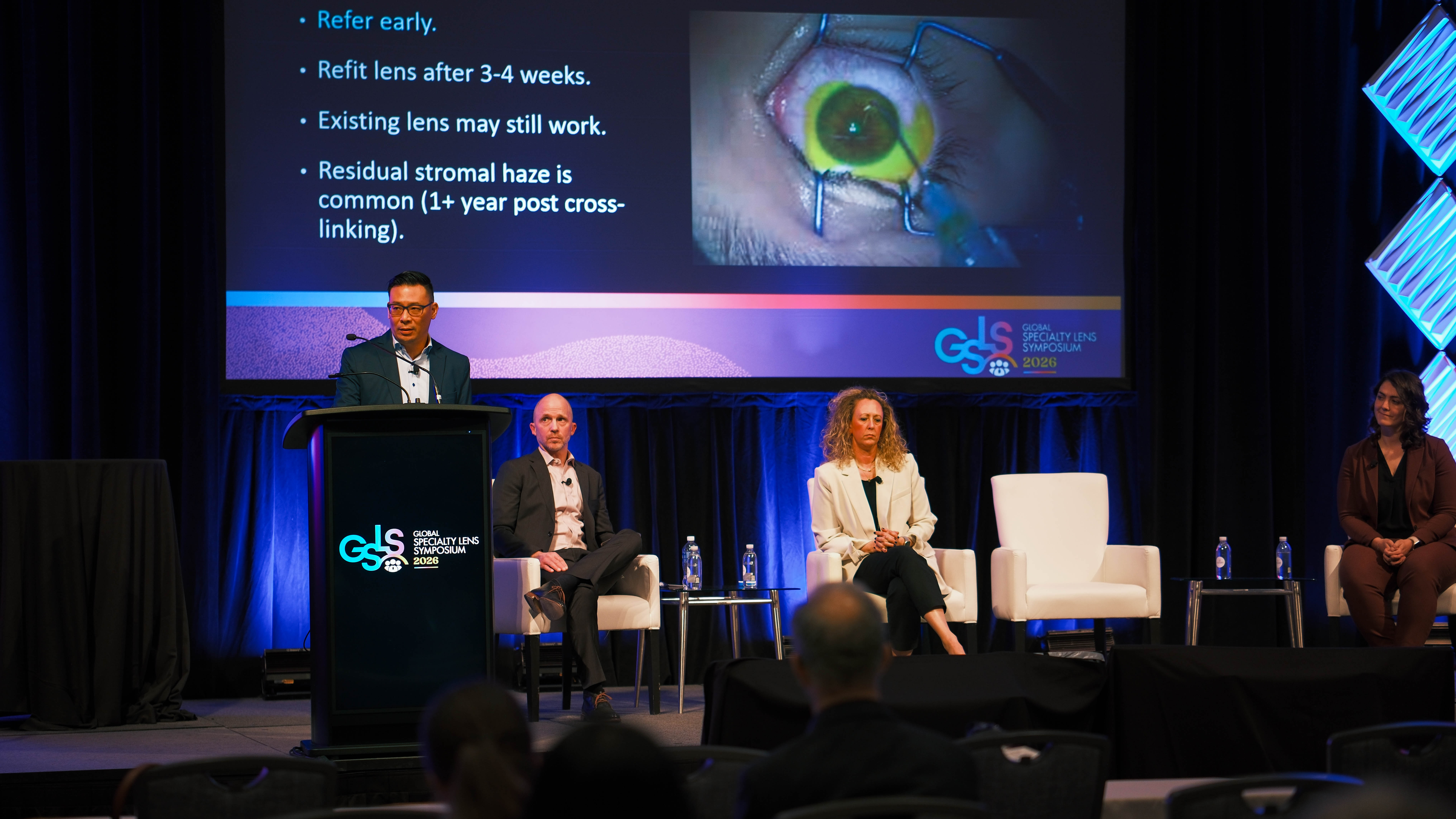
Clinical Report: Comprehensive Data Guides Keratoconus Crosslinking Decisions
Overview
A 15-year-old male with progressive keratoconus presented with severe thinning and steepening in the right eye and early signs in the left. Advanced imaging including epithelial mapping by OCT was critical to assess true stromal thickness and guide the decision for epithelium-off corneal crosslinking, highlighting the importance of comprehensive data in complex clinical scenarios.
Background
Keratoconus is a progressive corneal ectasia that can lead to significant visual impairment if untreated. Corneal crosslinking has revolutionized management by halting progression and reducing the need for transplantation. Accurate diagnosis and monitoring of progression are essential, typically relying on corneal tomography and clinical criteria. However, extreme corneal thinning and epithelial remodeling complicate assessment, necessitating advanced imaging techniques to safely guide treatment decisions.
Patient met progression criteria for corneal crosslinking in both eyes within 1 month, including steepening and thinning.
Right eye showed severe keratoconus with maximum keratometry of 83 D and significant corneal thinning and scarring.
OCT epithelial mapping revealed significant epithelial remodeling, reducing true stromal thickness to 262 µm in the right eye, below the safe threshold for crosslinking.
Corneal haze and scarring present preoperatively increase risk of post-crosslinking complications and visual acuity reduction.
Advanced imaging techniques are essential to accurately assess stromal thickness and epithelial changes, informing surgical eligibility and risk.
Clinical Implications
Clinicians should utilize comprehensive imaging, including epithelial mapping by OCT, when evaluating keratoconus patients with extreme corneal thinning to accurately determine candidacy for corneal crosslinking. Understanding epithelial remodeling and true stromal thickness is critical to avoid iatrogenic endothelial damage and to counsel patients effectively on risks and benefits. Early detection and routine monitoring remain key to preserving vision in keratoconus.
Conclusion
This case underscores the vital role of advanced diagnostic data in managing complex keratoconus cases, enabling safer and more informed clinical decisions regarding corneal crosslinking. Comprehensive assessment beyond traditional tomography is essential in the gray zones of clinical uncertainty.
References
Wollensak et al. 2003 -- Corneal Crosslinking: A New Treatment for Keratoconus
Koller et al. 2011 -- Criteria for Progression in Keratoconus
Hersh et al. 2017 -- Corneal Swelling Dynamics During Crosslinking
Mazzotta et al. 2018 -- Safety of Crosslinking in Thin Corneas
Reinstein et al. 2010 -- Epithelial Remodeling in Keratoconus
Wollensak et al. 2004 -- Corneal Haze After Crosslinking
Koller et al. 2014 -- Preoperative Densitometry Predicts Post-CXL Haze
This course discusses the diverse applications of scleral lenses beyond vision correction, including piggyback scleral lenses, oblate designs for keratoconus, scleral lenses for ptosis, drug delivery, prism correction, low vision, and optic decentration for fitting.