Clinical Report: Microglial Growth and Neuroinflammatory Changes Post-TBI
Overview
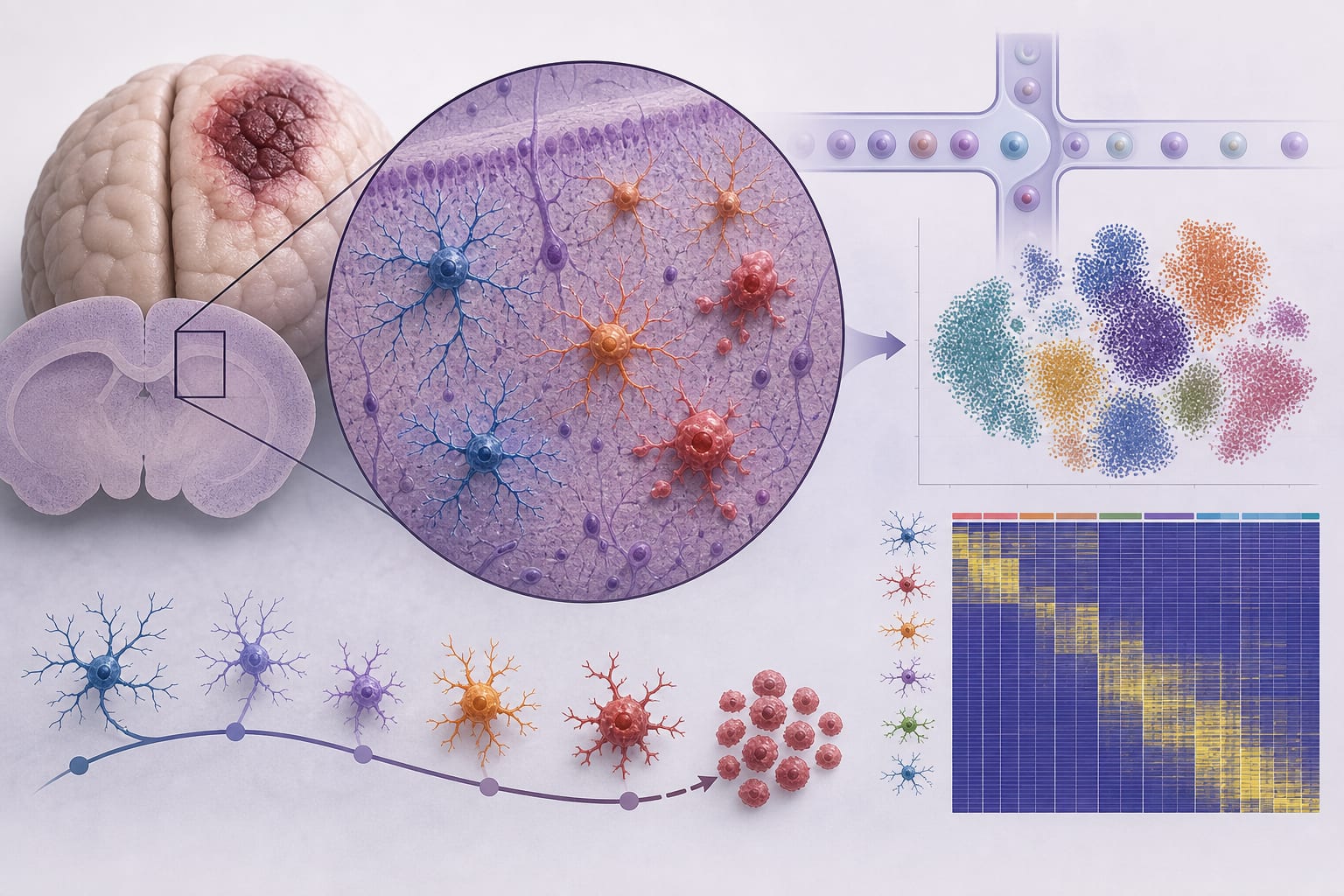
This study utilized single-cell RNA sequencing to analyze the transcriptional changes in cortical cells following traumatic brain injury (TBI) in a mouse model. Key findings include a significant reduction in homeostatic microglial subclusters and an increase in pro-inflammatory microglial phenotypes.
Background
Traumatic brain injury (TBI) is a significant global health issue, affecting millions annually and contributing to high mortality and disability rates. Understanding the cellular and molecular mechanisms of TBI is crucial. The neuroinflammatory response, primarily mediated by microglia, plays a central role in the secondary injury processes following TBI.
Data Highlights
No numerical data or trial data presented in the article.
Key Findings
Identification of 14 different cell groups in TBI and sham samples.
Reduction of homeostatic microglial subclusters (C0 and C2) after TBI.
Increased presence of disease-associated microglial subclusters (C1, C4, and C5) and a proliferation subgroup (MC Cycle).
Transition of microglial from a homeostatic state to a pro-inflammatory state post-TBI.
Enhanced intercellular communication and signaling pathways promoting M1 polarization in microglia.
Clinical Implications
The findings suggest that targeting the shift in microglial phenotypes may be a therapeutic strategy in managing neuroinflammation following TBI.
Conclusion
This study provides insights into the transcriptional changes in microglial populations following TBI.