Clinical Report: Meningococcal Abscess Detected by MRI
Overview
This case report describes an 11-year-old male with meningococcal meningitis who developed a cerebellar abscess detectable only by MRI. The patient's clinical condition improved significantly with targeted antimicrobial therapy and serial MRI monitoring.
Background
Meningococcal infections can lead to severe complications, including brain abscesses, which are rare in pediatric patients. Early detection and appropriate management are crucial for favorable outcomes. This case highlights the limitations of CT imaging in detecting early parenchymal infections, underscoring the importance of MRI in such scenarios.
Data Highlights
No numerical data or trial data was presented in the article.
Key Findings
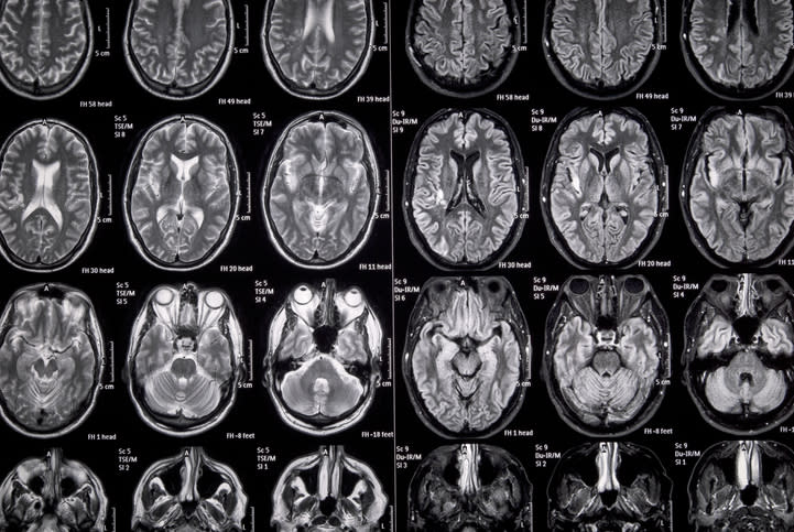
['The patient presented with fever, abdominal pain, and neurological decline.', 'Initial CT scan was unremarkable, but MRI revealed a cerebellar abscess.', 'Cerebrospinal fluid analysis showed 2,300 white blood cells per microliter with 98% neutrophils, elevated protein at 2.23 g/L, and low glucose at 0.67 mmol/L.', 'Polymerase chain reaction confirmed Neisseria meningitidis infection.', 'Serial MRI monitoring showed evolution of the abscess without mass effect.', 'The patient achieved full recovery with a 21-day course of intravenous ceftriaxone.']
Clinical Implications
Clinicians should maintain a high index of suspicion for complications in patients with meningitis, particularly when neurological symptoms persist or worsen. MRI should be considered for early detection of abscesses, especially when CT findings are inconclusive.
Conclusion
This case underscores the importance of early recognition and targeted therapy in managing meningococcal meningitis with complications. MRI plays a critical role in diagnosis and monitoring, leading to favorable outcomes without the need for surgical intervention.