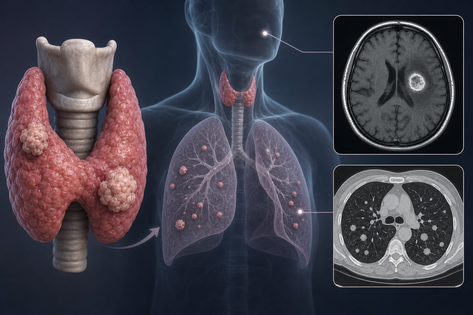
CT-Based Assessment of Malignancy Risk in Part-Solid Pulmonary Nodules
Overview
This study develops and validates CT-based models to predict malignancy in part-solid pulmonary nodules (PSNs). Incorporating qualitative vascular features significantly enhances predictive accuracy compared to traditional morphological assessments.
Background
The rise in lung cancer screening has led to increased detection of PSNs, which have a higher malignancy risk than pure ground-glass nodules. Accurate differentiation between malignant and benign PSNs is crucial for effective clinical management, as misclassification can lead to unnecessary procedures or delayed cancer diagnosis. Current guidelines primarily focus on nodule size and growth, highlighting the need for improved imaging biomarkers.
Data Highlights
Model
Training AUC
Testing AUC
Model 1
0.860
0.827
Model 2
0.916
0.898
Model 3
0.866
0.823
Key Findings
Malignant PSNs were associated with older age and female predominance.
Specific CT features such as irregular shape and spiculation were more common in malignant nodules.
Vascular patterns IV (interruption) and V (distortion) were significantly more prevalent in malignant nodules.
Model 2, which included vascular types IV and V, showed superior predictive performance with an AUC of 0.916 in training and 0.898 in testing.
Model 2 provided the highest net clinical benefit across various threshold probabilities.
Clinical Implications
Detail the potential impact of improved risk stratification on patient management.
Conclusion
The integration of vascular features into CT assessments for PSNs markedly improves diagnostic accuracy. This advancement has the potential to refine clinical management strategies for patients with pulmonary nodules.