Challenges in Segmenting Intraoperative Ultrasound Images for Brain Tumor Surgery
Overview
This study evaluates the difficulty in accurately delineating brain tumor boundaries on intraoperative ultrasound (iUS) images by measuring interobserver variation among experienced clinicians. It also models the clarity of tumor boundaries and assesses the potential of bounding box annotations as a simplified alternative to segmentation.
Background
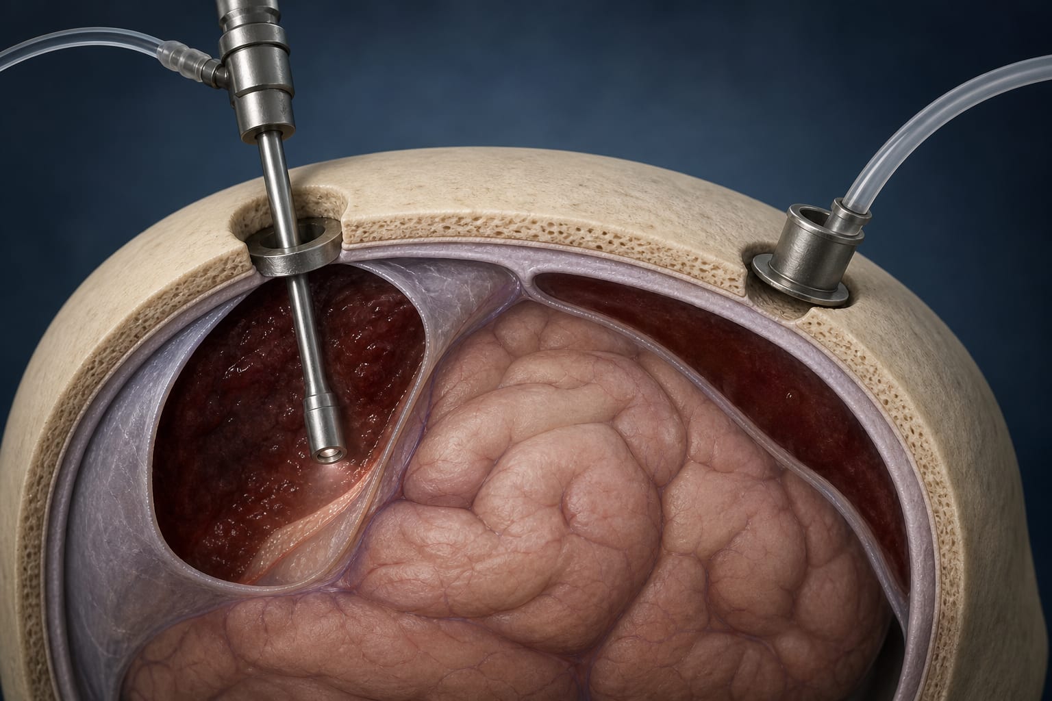
Maximal-safe resection is critical in brain tumor surgery to improve patient outcomes, and intraoperative ultrasound (iUS) offers real-time tumor visualization integrated into surgical workflow. Despite its advantages over intraoperative MRI, iUS adoption is limited by challenges including image quality, learning curve, and variability in tumor appearance. Accurate tumor boundary delineation on iUS remains difficult, impacting surgical precision and outcomes.
Data Highlights
The study involved 4 experienced annotators (1 neuroradiologist and 3 neurosurgeons) segmenting tumor boundaries on 30 iUS images from brain tumor surgeries. Images were acquired with a Canon i900 US machine and annotated using 3D Slicer software. The neuroradiologist's annotations, aided by full US datasets and preoperative MRI, served as the reference standard. Interobserver variation and pixel intensity modeling were used to assess segmentation clarity and consistency.
Key Findings
Significant interobserver variation exists in tumor boundary segmentation on iUS images among experienced clinicians.
Pixel intensity modeling revealed issues of low resolution and low signal-to-noise ratio contributing to boundary blurriness.
Bounding box annotations showed potential as a simpler, complementary method for outlining lesion margins.
Bounding boxes may help guide and improve segmentation accuracy by reducing uncertainty in tumor margin delineation.
The inherent variability of brain tumor appearances and intraoperative changes complicates consistent tumor boundary detection on iUS.
Clinical Implications
Understanding the limitations and variability in iUS tumor boundary delineation is essential for improving surgical guidance. Simplified annotation methods like bounding boxes could support clinicians in achieving more consistent tumor margin identification. Standardized training and development of new tools are needed to enhance iUS accuracy and facilitate its broader adoption in neurosurgery.
Conclusion
This study highlights fundamental challenges in segmenting brain tumor boundaries on intraoperative ultrasound images, emphasizing the need for improved techniques and training to reduce variability and enhance surgical outcomes. Bounding box annotations offer a promising adjunct to traditional segmentation approaches.
References
Imperial College NHS Trust London Study -- US-CNS: Multiparametric Advanced Ultrasound Imaging of the Central Nervous System Intraoperatively and Through Gaps in the Bone