Clinical Report: Uncommon Presentation of Ocular Giant Cell Arteritis
Overview
This report discusses a rare case of bilateral orbital inflammation in a 75-year-old male with Giant Cell Arteritis (GCA), highlighting diagnostic challenges and the importance of neuroimaging. The case underscores the need for increased awareness of atypical GCA presentations to prevent delays in treatment.
Background
Giant Cell Arteritis is a significant systemic vasculitis that can lead to severe complications, including vision loss. While typical presentations are well-documented, atypical manifestations, such as bilateral orbital inflammation, are less recognized and can complicate diagnosis and management. Understanding these presentations is crucial for timely intervention and improved patient outcomes.
Data Highlights
No numerical data or trial data presented in the article.
Key Findings
The patient presented with episodic vision loss and headaches, leading to a diagnosis of GCA.
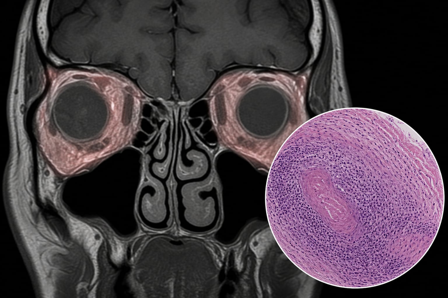
Neuroimaging revealed bilateral retrobulbar/perioptic soft-tissue infiltration, indicating an inflammatory process.
Temporal artery biopsy confirmed the diagnosis of temporal arteritis.
High-dose IV steroids initially improved symptoms, but the patient experienced persistent vision loss at follow-up.
Diagnostic challenges were noted due to the atypical presentation of GCA.
Current diagnostic criteria may need to incorporate advanced imaging techniques for better accuracy.
Clinical Implications
Clinicians should maintain a high index of suspicion for GCA in patients presenting with atypical ocular symptoms, particularly in older adults. Early neuroimaging and prompt initiation of glucocorticoids are critical to prevent irreversible vision loss.
Conclusion
This case emphasizes the importance of recognizing atypical presentations of GCA and the role of imaging in diagnosis. Continued research is needed to refine diagnostic criteria and improve management strategies for patients with GCA.