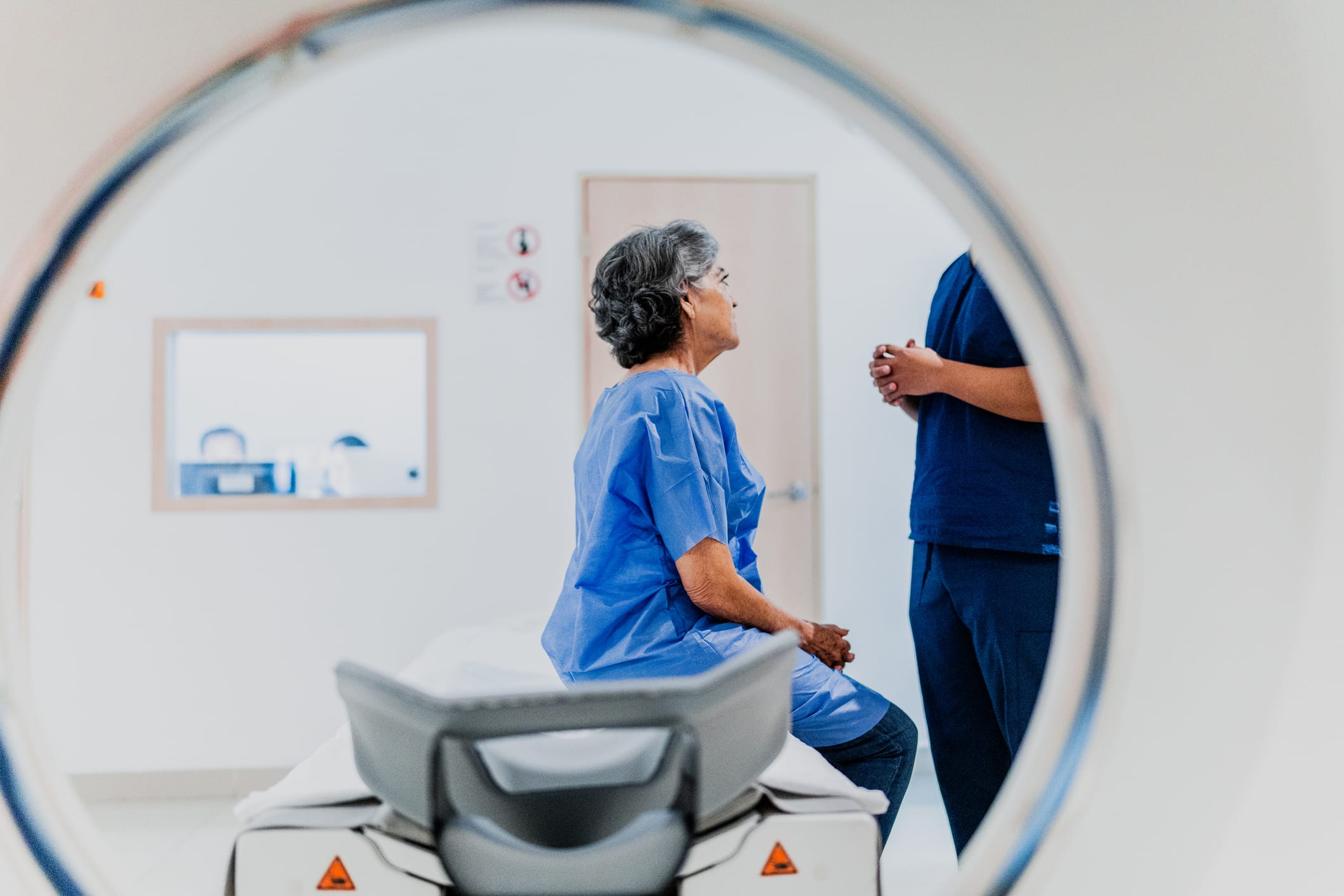
When to Choose MRN Over Ultrasound - Report - MDSpire
Advertisement
When to Choose MRN Over Ultrasound
Proximal location, multi-nerve involvement, and multi-region extent favor MRN; ultrasound retains value for dynamic assessment and metal artifact–limited cases.
Clinical Report: When to Choose MRN Over Ultrasound
Overview
Magnetic resonance neurography (MRN) demonstrates superior diagnostic value compared to high-resolution ultrasound (HRUS) in detecting upper-extremity peripheral neuropathies, particularly in complex cases, as evidenced by specific metrics such as odds ratios. This study highlights the importance of lesion topography in determining the choice of imaging modality.
Background
Upper-extremity peripheral neuropathies are common and can significantly impact patient quality of life. Accurate diagnosis is crucial for effective management, and advanced imaging techniques like MRN and HRUS play a vital role in this process. Understanding when to utilize each modality can enhance diagnostic accuracy and patient outcomes.
Data Highlights
Finding
MRN
HRUS
Concordant findings
60%
60%
Discordant findings
40%
40%
Cases with additional diagnostic value
261 (94.9%)
14 (5.1%)
Odds of added value (proximal lesions)
5.72 (95% CI: 3.42–9.56)
N/A
Odds of added value (multi-nerve disease)
1.65 (95% CI: 1.03–2.63)
N/A
Key Findings
MRN provided additional diagnostic value in 94.9% of cases compared to HRUS.
Proximal-only lesions had 5.72 times the odds of added diagnostic value from MRN.
Multi-anatomical-region involvement favored MRN with an odds ratio of 1.81.
Clinical symptoms were less predictive of MRN value than lesion anatomy.
HRUS was primarily useful for superficial focal lesions and dynamic assessments, but its effectiveness is highly operator-dependent.
Clinical Implications
Clinicians should consider MRN as the preferred imaging modality for complex upper-extremity neuropathies, especially when proximal lesions or multi-nerve involvement is suspected. HRUS may serve as a complementary tool in specific scenarios, particularly for superficial lesions, but its performance can vary significantly based on the operator's skill.
Conclusion
The findings support a tailored imaging approach, favoring MRN in complex cases while recognizing the utility of HRUS in certain contexts. Further studies are needed to validate these results across broader patient populations and different clinical settings.
Attenuation imaging increased with visually graded steatosis severity in pediatric patients, but findings were not validated against MRI-PDFF or biopsy.